
Snapping Hip Syndrome on MRI & X-ray: Imaging Findings
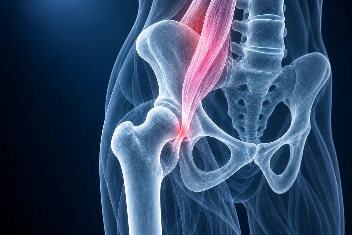
Snapping hip syndrome, also known as coxa saltans, is characterized by an audible or palpable snapping sensation during hip movement. The condition is classified as internal (most commonly the iliopsoas tendon snapping over the iliopectineal eminence or femoral head), external (the iliotibial band or gluteus maximus snapping over the greater trochanter), or intra-articular (caused by loose bodies, labral tears, or synovial chondromatosis). While often benign, persistent snapping hip can become painful and debilitating. MRI and dynamic ultrasound are key imaging tools for identifying the underlying anatomical cause.
Common Symptoms
- Audible or palpable snapping, clicking, or popping around the hip joint during movement
- External snapping over the greater trochanter caused by the iliotibial band flicking across the prominence during flexion-extension
- Internal snapping felt deep in the groin caused by the iliopsoas tendon catching on the iliopectineal eminence or femoral head
- Pain accompanying the snap, distinguishing pathological from benign snapping that is painless
- Worsening with repetitive hip flexion activities such as running, dance, or kicking
- Occasionally associated with lateral hip tenderness consistent with concurrent trochanteric bursitis
What We Look For on Imaging
- Iliopsoas tendon thickening or tendinopathy at the musculotendinous junction
- Iliopsoas bursitis with fluid distension adjacent to the hip joint
- Iliotibial band thickening or signal abnormality over the greater trochanter
- Trochanteric bursitis associated with external snapping
- Intra-articular loose bodies or synovial chondromatosis
- Associated labral tears or cartilage damage contributing to intra-articular snapping
Frequently Asked Questions
How is dynamic ultrasound used to diagnose snapping hip syndrome?
Dynamic ultrasound is the preferred imaging modality for snapping hip because it visualizes tendon movement in real time during provocation maneuvers. For external snapping hip, the transducer is placed over the greater trochanter while the patient performs hip flexion-extension; abrupt anterior displacement of the iliotibial band or posterior gluteus maximus edge across the trochanter is directly visualized. For internal snapping hip, the transducer is placed in the inguinal region during active hip flexion and extension from a position of external rotation; sudden medial-to-lateral displacement of the iliopsoas tendon over the iliopectineal eminence produces the characteristic snap. Associated peritendinous fluid, bursitis, or tendon thickening can be assessed simultaneously. MRI and MR arthrogram are reserved for evaluating intra-articular causes of snapping, including labral tears producing the symptom.
Can snapping hip cause labral tears or joint damage over time?
Internal snapping hip, specifically repetitive anterior displacement of the iliopsoas tendon, exerts compressive and shear forces on the anterior acetabular labrum at each snap. Chronic mechanical irritation can produce anterior labral tears, paralabral cysts, and chondral damage at the anterosuperior labrum, particularly in athletes with high-frequency repetitive hip flexion loads. MR arthrogram with radial reformats is used to detect associated labral pathology in patients with internal snapping hip and persistent pain that does not respond to conservative measures. Surgical treatment combining iliopsoas fractional lengthening with arthroscopic labral repair addresses both the mechanical cause and the consequent intra-articular damage.
What conservative treatments are effective for painful snapping hip?
Initial management prioritizes activity modification to reduce provocative movements, combined with a structured physical therapy program. For external snapping hip, therapy focuses on iliotibial band flexibility, hip abductor strengthening, and lumbopelvic control to reduce compressive loading at the trochanter. For internal snapping hip, progressive iliopsoas stretching and eccentric strengthening through functional range exercises are central. Corticosteroid injection into the iliopsoas bursa or the trochanteric bursa, performed under ultrasound guidance, can reduce inflammation and facilitate rehabilitation. Most patients with snapping hip improve with conservative care over 3 to 6 months; surgery is reserved for those with refractory symptoms or confirmed concomitant intra-articular pathology.
Sources
- Allen WC, Cope R. Coxa saltans: the snapping hip revisited. J Am Acad Orthop Surg. 1995;3(5):303-308.
- Lewis CL. Extra-articular snapping hip: a literature review. Sports Health. 2010;2(3):186-190.
- Deslandes M, Guillin R, Cardinal E, et al. The snapping iliopsoas tendon: new mechanisms using dynamic sonography. AJR Am J Roentgenol. 2008;190(3):576-581.
- Domb BG, Shindle MK, McArthur B, et al. Iliopsoas impingement: a newly identified cause of labral pathology in the hip. HSS J. 2011;7(2):145-150.
Understand your imaging before your next appointment
MRI, X-ray or CT from a CD, folder or ZIP. A PDF report or photos of the films work too.
Free to open and preview, no account needed. An optional AI report from $10 explains your images in plain language, with questions for your doctor.
Your raw files stay on this device. Only rendered images and minimal context are sent when you request an AI report.
Medical Disclaimer: This page is for informational and educational purposes only. It does not constitute medical advice, diagnosis, or treatment. AI-generated analysis may contain errors. Always consult a qualified healthcare professional for medical decisions. Full Disclaimer