
Hip Arthritis on X-ray: Imaging Findings
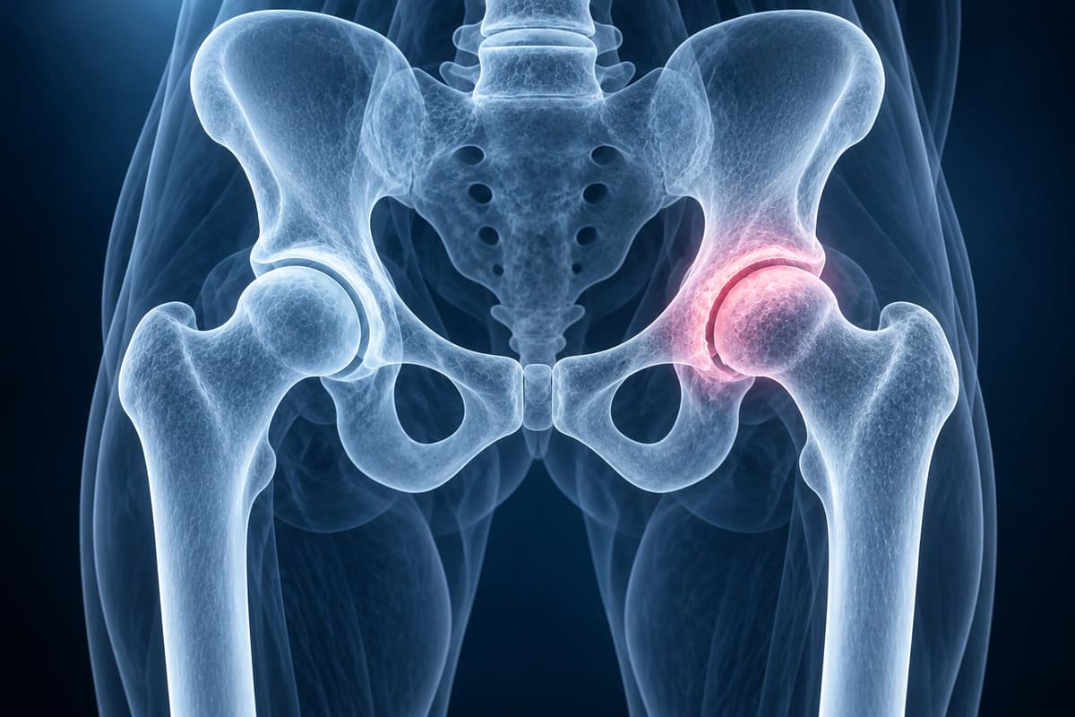
Hip osteoarthritis is a degenerative condition characterized by progressive loss of articular cartilage, osteophyte formation, and subchondral bone changes. It is a leading cause of disability and joint replacement surgery. Imaging assessment includes radiographic grading (Kellgren-Lawrence, Tonnis), measurement of joint space width, and MRI evaluation of cartilage status and bone marrow changes. AI can help explain arthritis grading terms, but a hip specialist must confirm the findings and discuss treatment or hip replacement planning.
Common Symptoms
- Deep groin, anterior thigh, or buttock pain that develops gradually and worsens with weight-bearing activity
- Morning stiffness lasting more than 30 minutes, improving with movement throughout the day
- Decreased internal rotation and hip flexion, making it difficult to tie shoes or enter a car
- Antalgic gait with a shortened stride on the affected side
- Night pain that disrupts sleep, particularly when lying on the affected hip
- Crepitus — a grinding or crunching sensation — palpable or audible during hip movement
Key Imaging Findings
- Joint space narrowing, typically superolateral in primary OA
- Marginal osteophyte formation at the femoral head-neck junction and acetabular rim
- Subchondral sclerosis and cyst formation
- Femoral head flattening or deformity in advanced disease
- Cartilage thinning and loss on MRI
- Bone marrow edema indicating active disease on MRI
Frequently Asked Questions
How is hip osteoarthritis graded on radiographs, and what do the grades mean for treatment?
The Tonnis grading system classifies hip osteoarthritis on AP pelvis radiographs. Grade 0 indicates no signs of arthritis. Grade 1 shows increased sclerosis of the femoral head or acetabulum, slight joint space narrowing, and minimal osteophyte formation. Grade 2 demonstrates small cysts, moderate joint space narrowing to less than 50% of normal, and moderate head deformity. Grade 3 reveals large cysts, severe joint space narrowing or obliteration, severe head deformity, and osteonecrosis. Treatment escalates with grade: grades 0–1 are managed conservatively, grade 2 may warrant joint-preserving procedures, and grade 3 typically necessitates total hip arthroplasty.
What does the lateral center-edge angle indicate in hip arthritis evaluation?
The lateral center-edge angle (LCEA), measured on an AP pelvis radiograph, quantifies acetabular coverage of the femoral head. A normal LCEA ranges from 25° to 39°. An LCEA below 25° indicates lateral uncoverage (hip dysplasia), which concentrates joint reaction forces on a reduced contact area and accelerates cartilage degeneration, producing early-onset arthritis in young adults. Conversely, an LCEA above 40° signals over-coverage associated with pincer FAI morphology. Both extremes predispose the hip to osteoarthritis through different mechanical pathways, and identifying acetabular morphology is essential for surgical planning in younger patients.
Can MRI detect hip arthritis earlier than radiographs?
Yes. Radiographic joint space narrowing reflects substantial cartilage loss that has already occurred, whereas MRI — especially T2 mapping and dGEMRIC (delayed gadolinium-enhanced MRI of cartilage) sequences — can detect biochemical changes in articular cartilage proteoglycan content before structural degradation becomes visible. Standard MRI also identifies early subchondral bone marrow edema, synovial hypertrophy, small effusions, and labral pathology in pre-arthritic hips. These findings help stratify patients who may benefit from joint-preserving surgery before irreversible cartilage loss narrows their treatment options.
Sources
- Kim YJ, Jaramillo D, Millis MB, et al. Assessment of early osteoarthritis in hip dysplasia with delayed gadolinium-enhanced magnetic resonance imaging of cartilage. J Bone Joint Surg Am. 2003;85(10):1987-1992.
- Domb BG, Gui C, Lodhia P. How much arthritis is too much for hip arthroscopy: a systematic review. Arthroscopy. 2015;31(3):520-529.
- Murphy NJ, Eyles JP, Hunter DJ. Hip Osteoarthritis: Etiopathogenesis and Implications for Management. Adv Ther. 2016;33(11):1921-1946.
Related Articles
Avascular necrosis of the femoral head — Ficat I-IV and ARCO 1-4 staging, MRI signs (double-line sign, crescent sign, collapse), and joint-preserving vs replacement options.
Understand your hip MRI report including labrum evaluation, cam and pincer morphology, cartilage assessment, and AVN detection.
Understand your imaging before your next appointment
MRI, X-ray or CT from a CD, folder or ZIP. A PDF report or photos of the films work too.
Free to open and preview, no account needed. An optional AI report from $10 explains your images in plain language, with questions for your doctor.
Your raw files stay on this device. Only rendered images and minimal context are sent when you request an AI report.
Medical Disclaimer: This page is for informational and educational purposes only. It does not constitute medical advice, diagnosis, or treatment. AI-generated analysis may contain errors. Always consult a qualified healthcare professional for medical decisions. Full Disclaimer