
Hip Impingement on MRI: Imaging Findings
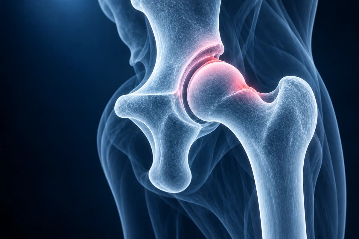
Femoroacetabular impingement (FAI) occurs when abnormal bony morphology of the femoral head (cam type), acetabulum (pincer type), or both (mixed type) causes abnormal contact during hip motion. This leads to labral tears, cartilage damage, and eventually osteoarthritis. Imaging is essential for identifying FAI morphology, measuring alpha angles, assessing acetabular coverage, and evaluating associated soft tissue damage. AI can help explain the shapes and measurements mentioned in your report, but a hip specialist must confirm them before any surgical planning.
Common Symptoms
- Sharp or aching groin pain that worsens with hip flexion, such as sitting for long periods or climbing stairs
- Pain at the end range of internal rotation and adduction of the hip
- Stiffness and reduced range of motion, particularly difficulty crossing the legs or putting on socks
- Clicking or catching sensation inside the hip joint during movement
- Pain during athletic activities involving pivoting, sprinting, or deep squatting
- Gradual onset buttock or lateral hip ache that may radiate to the anterior thigh
Key Imaging Findings
- Cam morphology with elevated alpha angle (greater than 55 degrees)
- Femoral head-neck junction bump or asphericity
- Pincer morphology with acetabular overcoverage (crossover sign)
- Coxa profunda or protrusio acetabuli
- Associated labral tear, most commonly anterosuperior
- Cartilage delamination at the chondrolabral junction
Frequently Asked Questions
What is the difference between cam and pincer FAI, and how is each identified on imaging?
Femoroacetabular impingement (FAI) has two morphological subtypes. Cam impingement arises from an aspherical femoral head-neck junction — a bony prominence that shears the acetabular labrum and cartilage during flexion. It is quantified on AP pelvis or Dunn-view radiographs by the alpha angle; an alpha angle greater than 55° is the accepted threshold for cam deformity. Pincer impingement results from focal or global acetabular over-coverage, trapping the labrum between the acetabular rim and the femoral neck. It is assessed on AP pelvis radiographs using the lateral center-edge angle (LCEA); an LCEA above 40° indicates coxa profunda or protrusio and suggests pincer morphology. Mixed FAI, combining both subtypes, is the most common presentation.
What does MRI add beyond plain radiographs in evaluating hip impingement?
Conventional MRI and, more definitively, MR arthrogram (MRA) with intra-articular gadolinium provide soft-tissue detail unavailable on radiographs. MRA improves labral tear detection sensitivity to over 90%, delineates cartilage delamination at the chondrolabral junction, and identifies paralabral cysts. Radial reformats — oblique MRI sequences acquired perpendicular to the femoral neck at multiple clock-face positions — map the full circumference of the head-neck junction, allowing precise measurement of the alpha angle in the plane of maximum cam deformity, which is typically at the 1–2 o'clock position on radial imaging.
When is surgery recommended over conservative management for FAI?
Conservative treatment — activity modification, physical therapy targeting hip rotator strengthening and lumbopelvic stability, and anti-inflammatory medication — is the first-line approach for 3 to 6 months. Arthroscopic or open surgical correction is considered when symptoms persist despite adequate conservative care, when MRA confirms a repairable labral tear contributing to mechanical symptoms, or when progressive chondral damage is documented. Early intervention before advanced joint space narrowing yields better outcomes, as significant osteoarthritis (Tonnis grade ≥ 2) is a relative contraindication to joint-preserving surgery.
Sources
- Ganz R, Parvizi J, Beck M, et al. Femoroacetabular impingement: a cause for osteoarthritis of the hip. Clin Orthop Relat Res. 2003;(417):112-120.
- Nötzli HP, Wyss TF, Stoecklin CH, et al. The contour of the femoral head-neck junction as a predictor for the risk of anterior impingement. J Bone Joint Surg Br. 2002;84(4):556-560.
- Beck M, Kalhor M, Leunig M, Ganz R. Hip morphology influences the pattern of damage to the acetabular cartilage: femoroacetabular impingement as a cause of early osteoarthritis of the hip. J Bone Joint Surg Br. 2005;87(7):1012-1018.
- Smith TO, Hilton G, Toms AP, et al. The diagnostic accuracy of acetabular labral tears using magnetic resonance imaging and magnetic resonance arthrography: a meta-analysis. Eur Radiol. 2011;21(4):863-874.
- Domb BG, Gui C, Lodhia P. How much arthritis is too much for hip arthroscopy: a systematic review. Arthroscopy. 2015;31(3):520-529.
Related Articles
Find out if hip labral tears can resolve without surgery, the role of physiotherapy, and surgical options when conservative care fails.
Post-operative hip labral repair rehabilitation guide including weight-bearing protocols, exercises, and return-to-activity milestones.
Post-arthroscopy hip rehabilitation guide covering protection, mobility, strengthening, and return-to-activity phases.
Understand your imaging before your next appointment
MRI, X-ray or CT from a CD, folder or ZIP. A PDF report or photos of the films work too.
Free to open and preview, no account needed. An optional AI report from $10 explains your images in plain language, with questions for your doctor.
Your raw files stay on this device. Only rendered images and minimal context are sent when you request an AI report.
Medical Disclaimer: This page is for informational and educational purposes only. It does not constitute medical advice, diagnosis, or treatment. AI-generated analysis may contain errors. Always consult a qualified healthcare professional for medical decisions. Full Disclaimer