
Avascular Necrosis on MRI: Imaging Findings
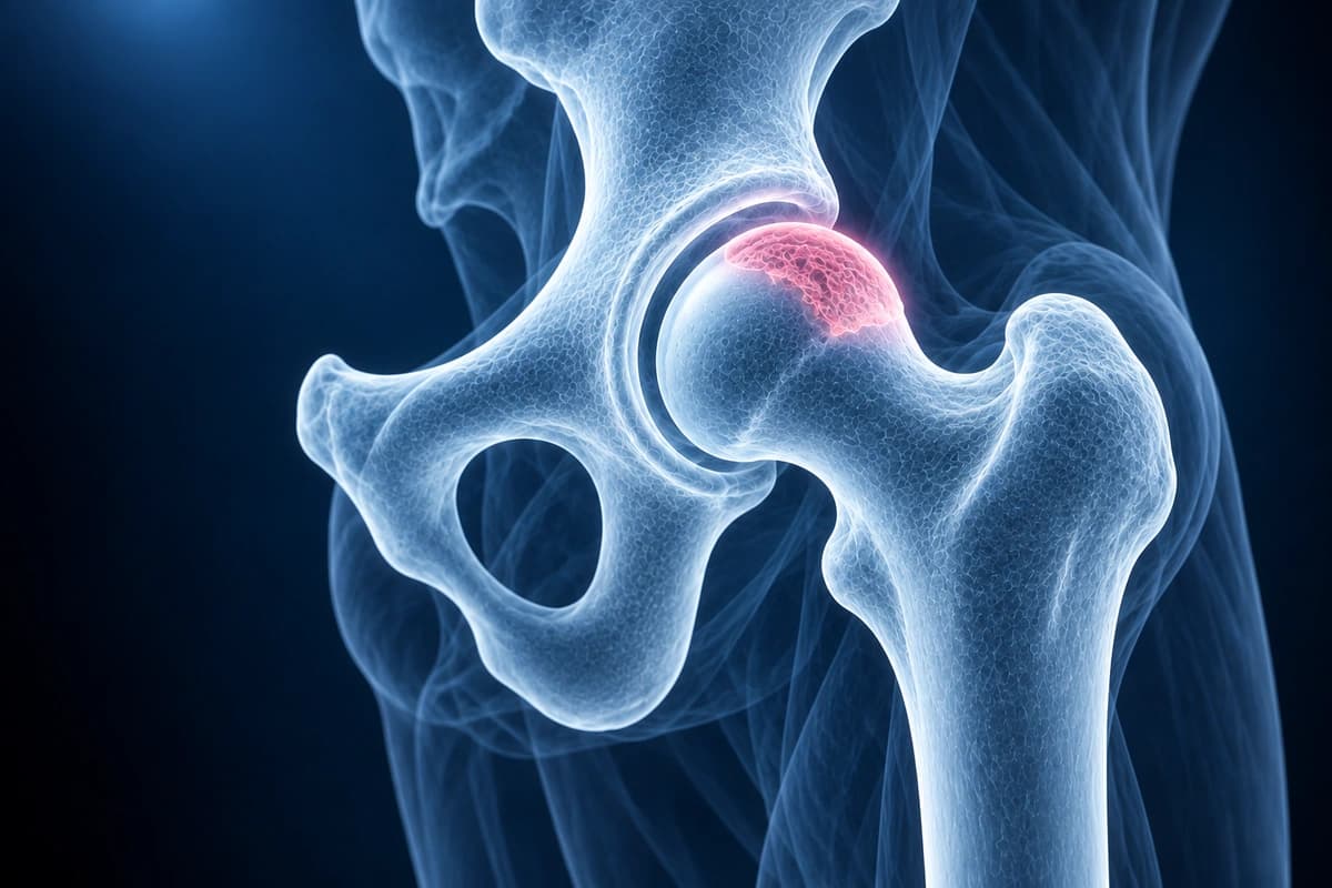
Avascular necrosis (AVN), also known as osteonecrosis, of the femoral head results from disruption of blood supply to the bone, leading to bone cell death and potential femoral head collapse. Early detection with MRI is critical for treatment planning and joint preservation. Risk factors include corticosteroid use, alcohol abuse, trauma, and sickle cell disease.
Common Symptoms
- Sudden or insidious onset of deep groin pain, often initially present only with weight-bearing and progressing to rest pain
- Pain that may precede any radiographic abnormality by weeks to months
- Antalgic gait developing as the femoral head collapses and joint congruity is lost
- Restricted hip range of motion, particularly internal rotation and abduction
- Bilateral hip involvement in up to 80% of cases associated with corticosteroid use or systemic disease
- Night pain and progressive inability to bear weight as the condition advances
Key Imaging Findings
- Band-like low signal on T1 in the femoral head (reactive interface)
- Double-line sign on T2 (pathognomonic for AVN)
- Bone marrow edema surrounding the necrotic segment
- Subchondral fracture (crescent sign) indicating early collapse
- Femoral head flattening or collapse in advanced disease
- Contralateral hip involvement assessment
Frequently Asked Questions
What is the AVN crescent sign, and what does it mean clinically?
The crescent sign is a subchondral radiolucent line visible on AP or frog-leg lateral hip radiographs, representing separation of the necrotic subchondral bone from the overlying articular cartilage. It indicates mechanical failure of the subchondral plate and imminent or actual femoral head collapse. Its presence upgrades the patient to at least Ficat stage III and dramatically changes prognosis — once the crescent sign appears, collapse typically occurs within weeks to months without intervention. Identifying the crescent sign on radiographs is therefore a critical triage point that often prompts urgent surgical consultation.
What does Ficat staging describe, and how is MRI used alongside it?
Early osteonecrosis may have normal radiographs despite MRI changes. Femoral head collapse marks progression beyond precollapse disease and changes treatment options.
Which risk factors most commonly cause avascular necrosis of the femoral head?
The most frequent causes are corticosteroid use (cumulative doses above 2 g of prednisone equivalent or high-dose pulse therapy) and excessive alcohol consumption, together accounting for roughly 90% of non-traumatic AVN cases. Traumatic AVN results from displaced femoral neck fractures or hip dislocations that disrupt the retinacular blood supply to the femoral head. Additional risk factors include systemic lupus erythematosus, sickle cell disease, hyperlipidemia, Gaucher disease, radiation therapy, and decompression sickness (dysbaric osteonecrosis). Idiopathic AVN accounts for a minority of cases. Recognizing the underlying etiology guides treatment of the precipitating condition alongside hip-specific management.
Sources
- Mitchell DG, Rao VM, Dalinka MK, et al. Femoral head avascular necrosis: correlation of MR imaging, radiographic staging, radionuclide imaging, and clinical findings. Radiology. 1987;162(3):709-715.
- Mont MA, Cherian JJ, Sierra RJ, et al. Nontraumatic osteonecrosis of the femoral head: where do we stand today? A ten-year update. J Bone Joint Surg Am. 2015;97(19):1604-1627.
- Moya-Angeler J, Gianakos AL, Villa JC, et al. Current concepts on osteonecrosis of the femoral head. World J Orthop. 2015;6(8):590-601.
Related Articles
Avascular necrosis of the femoral head — Ficat I-IV and ARCO 1-4 staging, MRI signs (double-line sign, crescent sign, collapse), and joint-preserving vs replacement options.
Learn about common hip conditions including labral tears, femoroacetabular impingement, bursitis, stress fractures, and avascular necrosis.
Learn when 3T hip MRI can help for labral tears, FAI, cartilage, stress fractures, and avascular necrosis, and when X-ray, 1.5T MRI, or MR arthrogram is enough.
Understand your imaging before your next appointment
MRI, X-ray or CT from a CD, folder or ZIP. A PDF report or photos of the films work too.
Free to open and preview, no account needed. An optional AI report from $10 explains your images in plain language, with questions for your doctor.
Your raw files stay on this device. Only rendered images and minimal context are sent when you request an AI report.
Medical Disclaimer: This page is for informational and educational purposes only. It does not constitute medical advice, diagnosis, or treatment. AI-generated analysis may contain errors. Always consult a qualified healthcare professional for medical decisions. Full Disclaimer