
Hip Fracture on X-ray: Imaging Findings
Hip fractures are serious injuries, particularly in elderly patients with osteoporosis. They are classified as intracapsular (femoral neck) or extracapsular (intertrochanteric, subtrochanteric). Classification guides treatment: displaced femoral neck fractures often require arthroplasty, while intertrochanteric fractures are typically fixed with intramedullary devices. MRI is highly sensitive for detecting occult fractures not visible on initial X-rays.
Some signs described on this page need prompt medical care. If you have them, get care first. Reading about imaging can wait.
Common Symptoms
- Acute severe groin or proximal thigh pain following a fall or, in osteoporotic bone, minimal trauma
- Inability to bear weight on the affected limb immediately after injury
- Shortened and externally rotated lower extremity on inspection, most prominent in displaced fractures
- Pain with any attempted passive hip rotation, even in undisplaced fractures
- Marked tenderness on palpation of the greater trochanter, groin, or proximal femur
- In stress or insufficiency fractures, insidious onset groin pain that worsens progressively with activity without a clear traumatic event
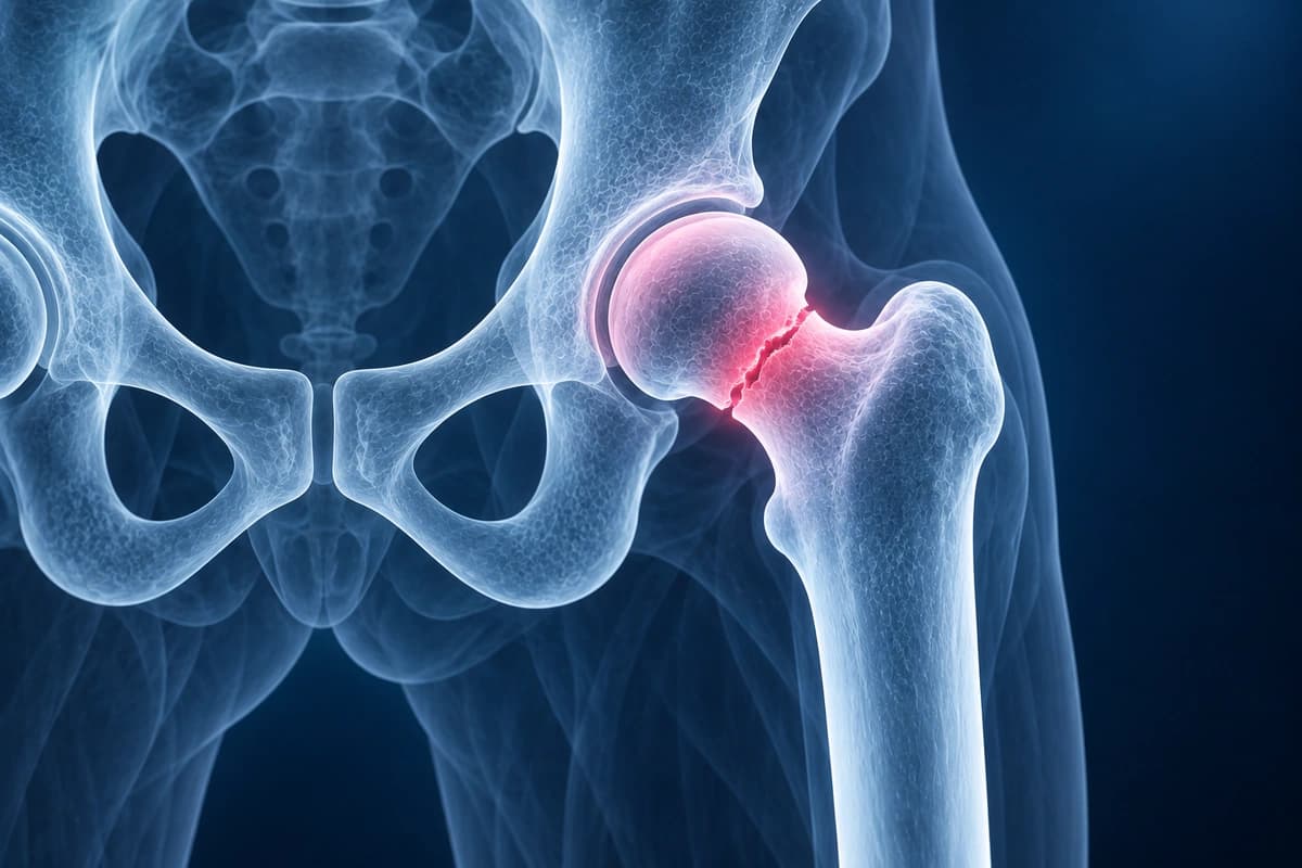
Key Imaging Findings
- Fracture line through the femoral neck with Garden classification
- Intertrochanteric fracture pattern with fragment assessment
- Subtrochanteric fracture extension
- Bone marrow edema on MRI indicating occult fracture
- Displacement and angulation measurement
- Stress fracture or insufficiency fracture on MRI not visible on X-ray
Frequently Asked Questions
How are hip fractures classified and why does classification matter for treatment?
Hip fractures are divided anatomically into femoral neck fractures and intertrochanteric or subtrochanteric fractures. Femoral neck fractures are further classified by the Garden classification (stages I–IV based on displacement) or the Pauwels classification (types I–III based on fracture line angle). Displacement is the primary driver of treatment: non-displaced femoral neck fractures (Garden I–II) in physiologically young patients are managed with internal fixation using cannulated screws or dynamic hip screws, whereas displaced fractures carry high avascular necrosis and non-union rates, prompting hemiarthroplasty or total hip arthroplasty in most patients over 65. Intertrochanteric fractures, which have a more robust blood supply, are almost universally treated with fixation using a sliding hip screw or intramedullary nail.
When is MRI preferred over CT for diagnosing suspected hip fracture?
A meta-analysis estimated CT sensitivity at 94% for occult proximal femoral fractures. Persistent clinical concern after a normal CT should prompt MRI.
What is the significance of timing to surgery in hip fracture management?
Evidence consistently shows that surgical fixation or arthroplasty performed within 24 to 48 hours of admission is associated with lower rates of mortality, pulmonary embolism, pressure ulcers, and pneumonia compared with delayed surgery. Prolonged bed rest in elderly patients accelerates deconditioning and increases thromboembolic risk despite anticoagulation. Medical optimization — addressing anemia, anticoagulant reversal, cardiac evaluation, and electrolyte correction — should proceed rapidly in parallel with surgical planning rather than serially. Hip fracture programs with dedicated pathways targeting surgery within 24 hours have demonstrated meaningful reductions in 30-day and 1-year mortality across multiple health systems.
Sources
- Kellock TT, Khurana B, Mandell JC. Diagnostic performance of CT for occult proximal femoral fractures: a systematic review and meta-analysis. AJR Am J Roentgenol. 2019;213(6):1324-1330.
- Expert Panel on Musculoskeletal Imaging; Ross AB, Lee KS, et al. ACR Appropriateness Criteria Acute Hip Pain-Suspected Fracture. J Am Coll Radiol. 2019;16(5S):S18-S25.
- Moja L, Piatti A, Pecoraro V, et al. Timing matters in hip fracture surgery: patients operated within 48 hours have better outcomes. A meta-analysis and meta-regression of over 190,000 patients. PLoS One. 2012;7(10):e46175.
Do not delay urgent medical care to use this tool.
After you get care, understand your imaging
MRI, X-ray or CT from a CD, folder or ZIP. A PDF report or photos of the films work too.
Free to open and preview, no account needed. An optional AI report from $10 explains your images in plain language, with questions for your doctor.
Your raw files stay on this device. Only rendered images and minimal context are sent when you request an AI report.
Medical Disclaimer: This page is for informational and educational purposes only. It does not constitute medical advice, diagnosis, or treatment. AI-generated analysis may contain errors. Always consult a qualified healthcare professional for medical decisions. Full Disclaimer