
Common Symptoms
- Lateral hip pain centred over the greater trochanter
- Pain when lying on the affected side at night
- Pain with single-leg stance lasting more than 30 seconds
- Trendelenburg gait or hip drop when walking
- Pain when climbing stairs or rising from a low chair
- Tenderness directly over the greater trochanter on palpation
Gluteal Tendinopathy on MRI: AI-Powered Imaging Analysis
AI-powered gluteal tendinopathy and greater trochanteric pain syndrome analysis on hip MRI. Understand gluteus medius and minimus tears, trochanteric bursitis, and tendon thickening.
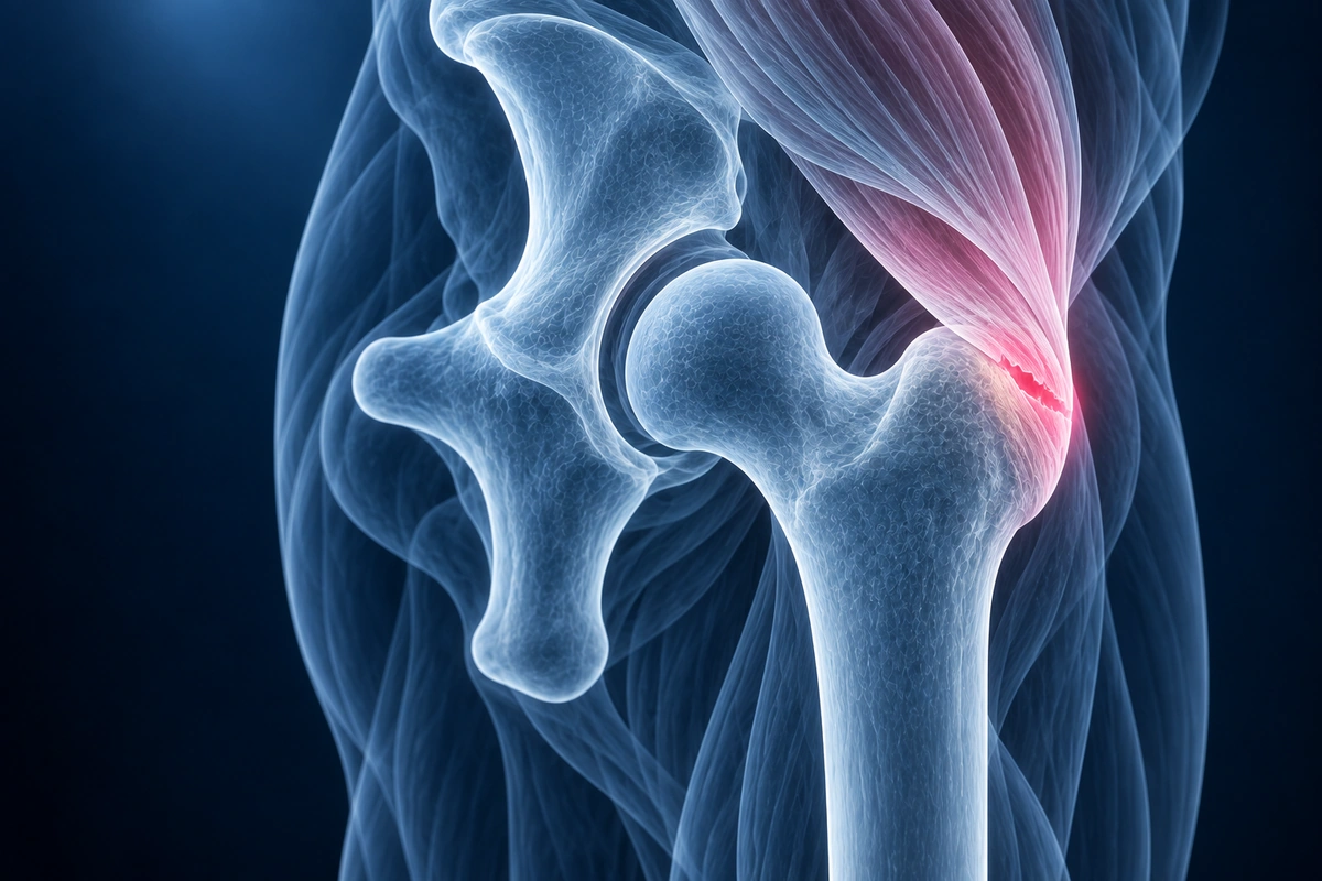
Gluteal tendinopathy — pathology of the gluteus medius and gluteus minimus tendons at their insertion on the greater trochanter — is the modern, evidence-based explanation for what was historically labelled trochanteric bursitis. It affects approximately 25% of middle-aged women and is among the most common causes of lateral hip pain, yet it is frequently misdiagnosed or dismissed. Trochanteric bursitis, when present, is almost always a secondary finding caused by the overlying tendon disease irritating the bursa — treating only the bursa without addressing the tendon pathology leads to disappointingly high recurrence rates. MRI is the gold standard for distinguishing tendon thickening, partial tearing, and complete tearing, and for confirming whether bursal involvement is truly primary or secondary.
What We Look For on Imaging
- Gluteus medius or minimus tendon thickening with increased signal on T2 fat-saturated sequences at the greater trochanter insertion
- Partial or complete tearing of the gluteal tendon footprint, seen as a fluid-signal gap within or deep to the tendon
- Peritendinous oedema surrounding the gluteal tendon on coronal fat-suppressed images
- Secondary trochanteric bursal fluid — distension of the bursa superficial to the greater trochanter as a consequence of tendon pathology
- Greater trochanter cortical irregularity or enthesophyte formation at the tendon insertion site
- Assessment for intra-articular pathology — labral tears and hip joint effusion — that can refer pain laterally and mimic gluteal tendinopathy
Frequently Asked Questions
Is this bursitis or tendinopathy — and does it matter?
In the overwhelming majority of cases what patients are told is trochanteric bursitis is actually gluteal tendinopathy. High-quality MRI studies show that isolated primary bursitis without tendon involvement is uncommon; the tendon is almost always the primary problem. This distinction matters for treatment: corticosteroid injections into the bursa provide short-term relief in both conditions, but if the tendon is not rehabilitated, symptoms return. Correctly identifying tendinopathy on MRI directs treatment toward the tendon, which is where lasting improvement comes from.
Do I need surgery for gluteal tendinopathy?
Surgery is rarely needed. The vast majority of patients respond to well-structured non-operative management over 3 to 6 months. Surgery — typically arthroscopic tendon repair and bursectomy — is reserved for patients with confirmed full-thickness gluteal tendon tears who have failed a comprehensive conservative programme. Partial tears almost never require surgical repair. The decision to operate depends on the degree of tendon disruption on MRI, functional limitations, and the patient's response to non-surgical treatment.
What is the best treatment for gluteal tendinopathy?
The most effective evidence-based treatment is load management combined with a progressive strengthening programme targeting the gluteus medius and minimus. Patients are advised to avoid positions that compress the tendon — crossing the legs, sitting on low chairs, and sleeping with the hip adducted — because compressive load at the trochanteric insertion is a key driver of tendinopathy. Eccentric and isometric strengthening exercises, progressed under physiotherapist guidance, restore tendon capacity and reduce pain. Weight loss, where relevant, reduces load on the tendon with every step. For patients who do not improve adequately with exercise, extracorporeal shockwave therapy has good evidence for gluteal tendinopathy, and platelet-rich plasma (PRP) injection is an emerging option with promising early trial results. Corticosteroid injections provide faster short-term pain relief but do not address the underlying tendon problem and may cause tendon weakening with repeated use.
Upload your imaging to check for Gluteal Tendinopathy
Upload your MRI or X-ray DICOM files for private, AI-powered analysis. 4 models analyze independently — raw scan files stay in your browser.
Upload & AnalyzeMedical Disclaimer: This page is for informational and educational purposes only. It does not constitute medical advice, diagnosis, or treatment. AI-generated analysis may contain errors. Always consult a qualified healthcare professional for medical decisions. Full Disclaimer