
How to Read a Hip MRI: A Patient's Guide
Understand your hip MRI report including labrum evaluation, cam and pincer morphology, cartilage assessment, and AVN detection.
Key Takeaways
- MR arthrogram is the most sensitive technique for detecting hip labral tears
- Cam and pincer morphology on MRI helps identify the structural cause of impingement
- Paralabral cysts are strong indirect evidence of an underlying labral tear
- AVN shows a characteristic serpiginous line pattern on T1 images distinguishing it from other bone pathology
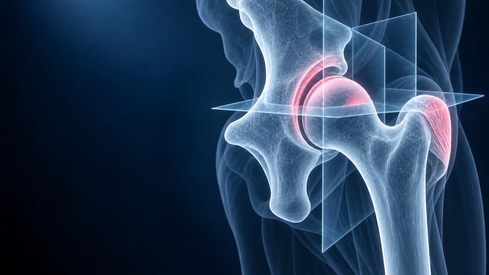
Hip MRI provides detailed visualization of the labrum, articular cartilage, bone morphology, tendons, and bursae that cannot be seen on X-rays. Whether your MRI was performed with or without arthrogram, understanding the key structures and common findings helps you participate in informed discussions about your treatment plan.
Standard MRI Sequences
- T1-weighted images: show bone anatomy and marrow detail, with normal fatty marrow appearing bright
- T2-weighted with fat saturation: highlight fluid, edema, and inflammation as bright signal against dark suppressed fat
- Proton density sequences: excellent for evaluating the labrum, cartilage, and ligamentum teres
- For acetabular labral tears, a meta-analysis estimated MR arthrography sensitivity at 87%. It can help evaluate the labrum, but it is not a perfect test.
Labrum on MRI
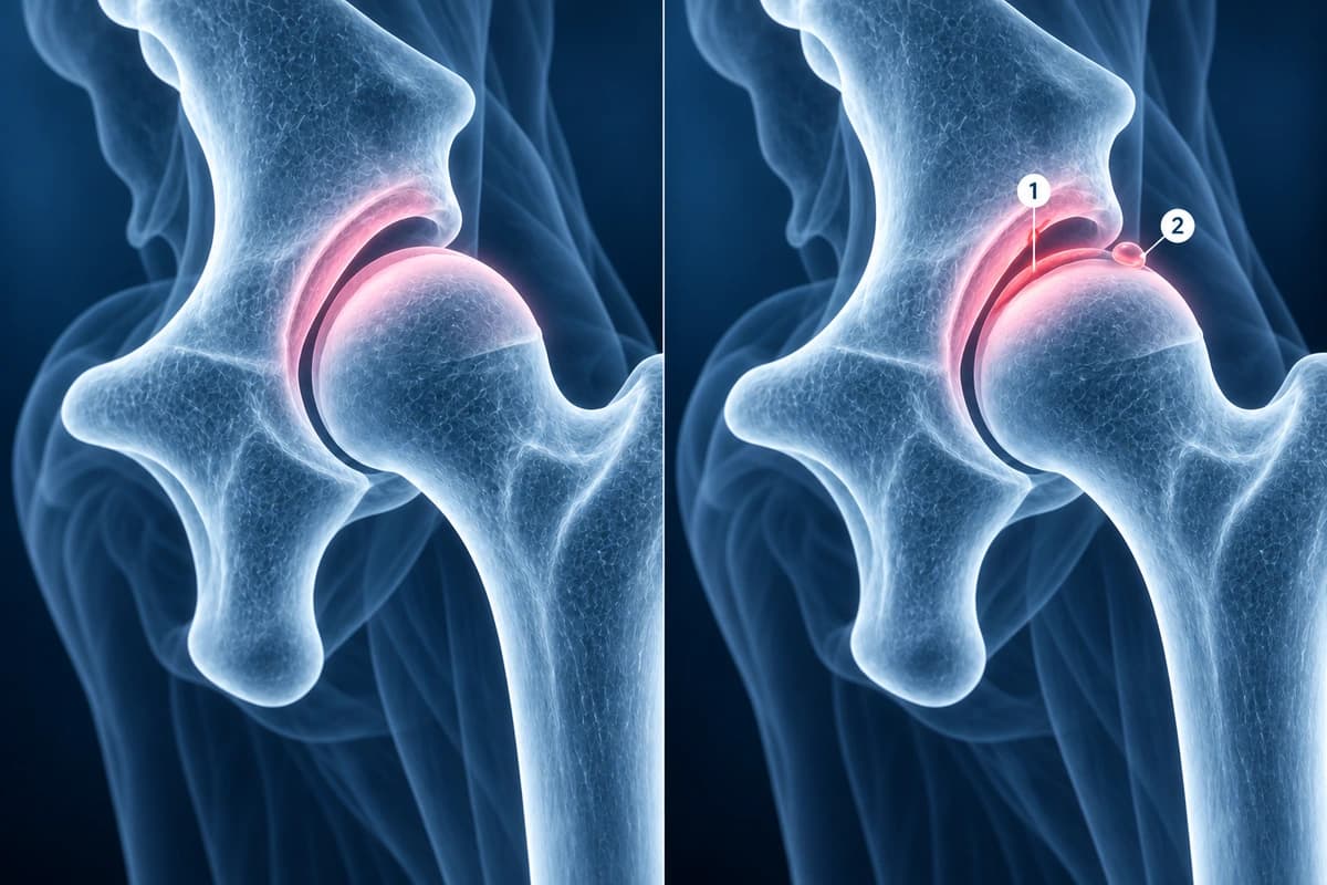
The normal labrum appears as a dark, triangular structure attached to the acetabular rim on coronal and sagittal images. Labral tears show as bright signal within or at the base of the labrum on T2 or arthrogram images. Tear patterns include detachment from the acetabular rim, intrasubstance degeneration, and flap tears. The anterior-superior labrum is the most commonly affected location. Paralabral cysts, which appear as bright fluid-filled structures adjacent to the labrum, are strong indirect signs of an underlying tear. For treatment options, see our article on labral tear healing without surgery.
Cam and Pincer Morphology
Femoroacetabular impingement morphology is assessed on MRI and may also be visible on X-rays. Cam morphology appears as a bump or asphericity at the femoral head-neck junction, measured by the alpha angle (greater than 55 degrees is abnormal). Pincer morphology shows as excessive acetabular coverage, measured by the lateral center-edge angle. Both types cause abnormal contact during hip motion, leading to labral tears and cartilage damage. For more on FAI and related conditions, see our hip injury types guide.
Cartilage and Bone Assessment
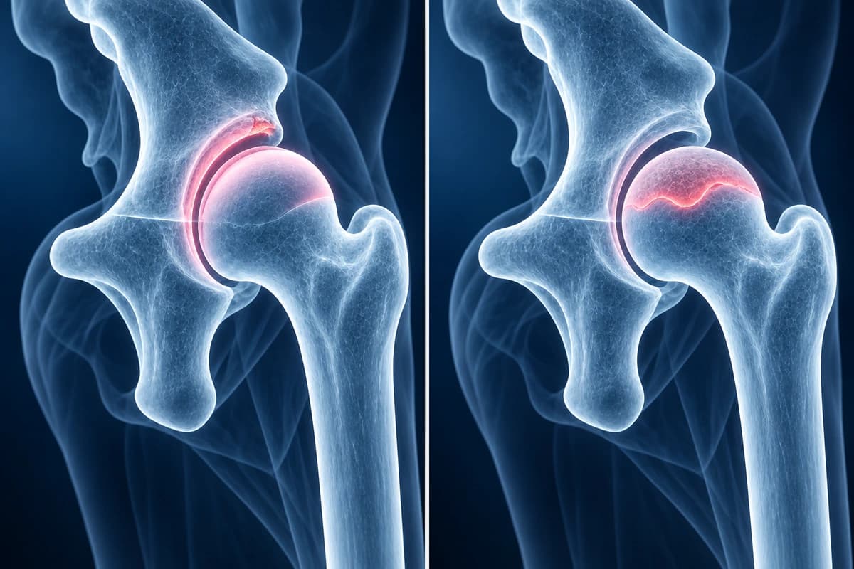
Articular cartilage appears as an intermediate-signal layer covering the femoral head and acetabulum. Cartilage damage shows as thinning, fissuring, or focal defects, often in the anterosuperior acetabulum where impingement forces concentrate. Bone marrow edema appears as bright T2/STIR signal within normally dark bone marrow and may indicate stress fractures, AVN, or transient osteoporosis. AVN specifically shows a characteristic serpiginous (wavy line) pattern of low signal on T1 images demarcating viable from necrotic bone.
Frequently Asked Questions
Do I need MR arthrogram or standard MRI for my hip?
MR arthrogram is preferred when labral tears are the primary concern, as the injected contrast outlines the labrum and significantly improves sensitivity. Standard MRI without arthrogram is sufficient for evaluating bone marrow pathology (AVN, stress fractures), tendon injuries, bursitis, and muscle conditions. Your orthopedic surgeon will typically specify which study is needed based on your clinical presentation.
Can hip MRI show early arthritis?
MRI can detect cartilage damage and early osteoarthritic changes before they become visible on X-rays. However, for established osteoarthritis, weight-bearing X-rays remain the primary imaging study because they show joint space narrowing under load, which MRI cannot replicate since the patient lies down during the scan.
Sources
- Seldes RM, Tan V, Hunt J, et al. Anatomy, histologic features, and vascularity of the adult acetabular labrum. Clin Orthop Relat Res. 2001;(382):232-240.
- Huang Z, Liu W, Li T, et al. Diagnostic value of magnetic resonance imaging and magnetic resonance arthrography for assessing acetabular labral tears: a systematic review and meta-analysis. Medicine (Baltimore). 2023;102(9):e32963.
- Nötzli HP, Wyss TF, Stoecklin CH, et al. The contour of the femoral head-neck junction as a predictor for the risk of anterior impingement. J Bone Joint Surg Br. 2002;84(4):556-560.
- Mitchell DG, Rao VM, Dalinka MK, et al. Femoral head avascular necrosis: correlation of MR imaging, radiographic staging, radionuclide imaging, and clinical findings. Radiology. 1987;162(3):709-715.
- Expert Panel on Musculoskeletal Imaging; Jawetz ST, Fox MG, et al. ACR Appropriateness Criteria Chronic Hip Pain: 2022 Update. J Am Coll Radiol. 2023;20(5S):S33-S48.
Related Articles
Learn about common hip conditions including labral tears, femoroacetabular impingement, bursitis, stress fractures, and avascular necrosis.
Find out if hip labral tears can resolve without surgery, the role of physiotherapy, and surgical options when conservative care fails.
Cam, pincer, and mixed femoroacetabular impingement on MRI — alpha angle, lateral center-edge angle, labral tear patterns, and treatment differences.
Related Conditions
Understand your imaging before your next appointment
MRI, X-ray or CT from a CD, folder or ZIP. A PDF report or photos of the films work too.
Free to open and preview, no account needed. An optional AI report from $10 explains your images in plain language, with questions for your doctor.
Your raw files stay on this device. Only rendered images and minimal context are sent when you request an AI report.
Medical Disclaimer: This page is for informational and educational purposes only. It does not constitute medical advice, diagnosis, or treatment. AI-generated analysis may contain errors. Always consult a qualified healthcare professional for medical decisions. Full Disclaimer