
Hip Stress Fracture on MRI & X-ray: Imaging Findings
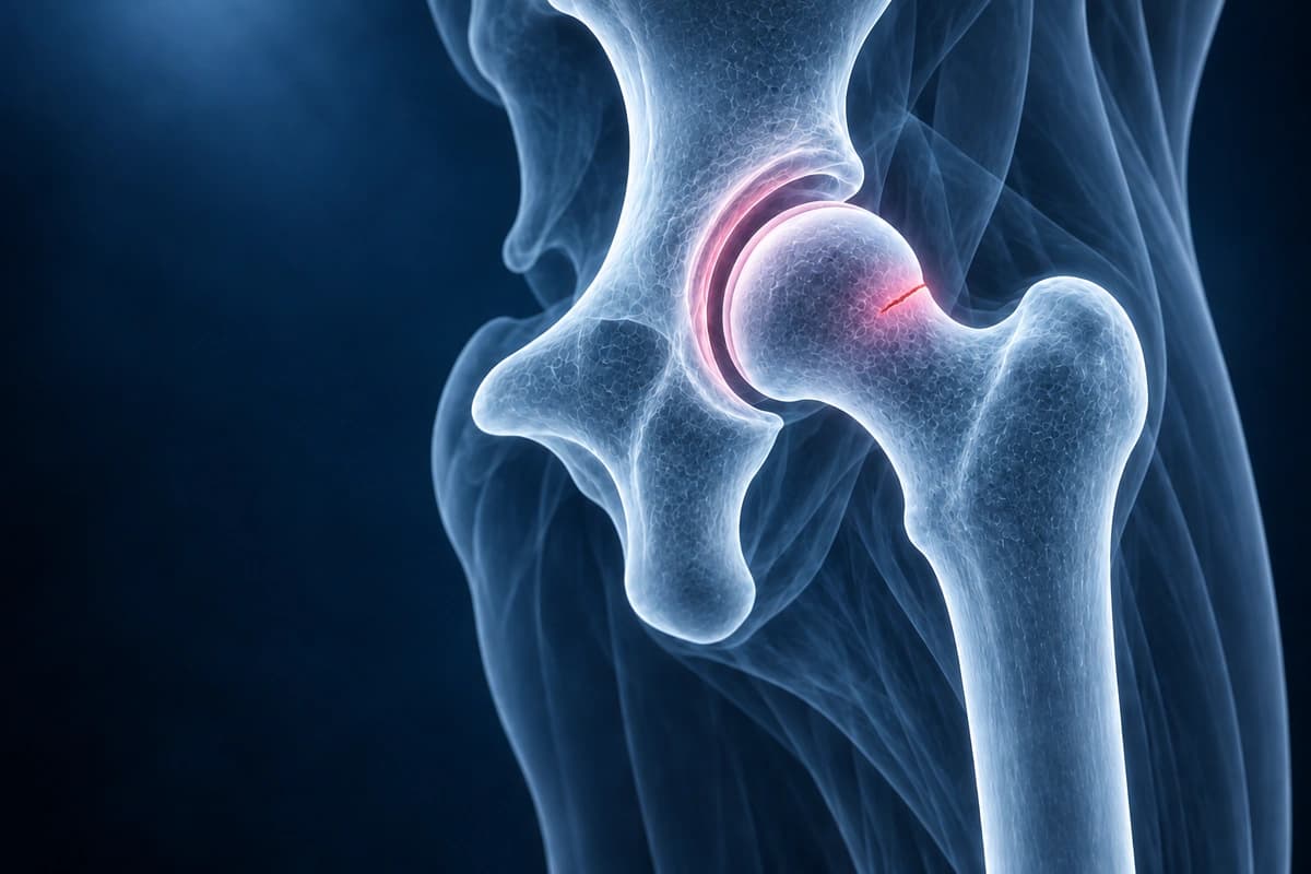
A hip stress fracture, most commonly involving the femoral neck, is an incomplete fracture caused by repetitive microtrauma that exceeds the bone's ability to remodel and repair. It is frequently seen in distance runners, military recruits, and athletes with relative energy deficiency in sport (RED-S). Femoral neck stress fractures are particularly concerning because they carry a significant risk of completion to a displaced fracture, which can compromise the femoral head blood supply and lead to avascular necrosis. MRI is the most sensitive imaging modality for early detection, often revealing bone marrow edema before a fracture line is visible on X-ray. AI can help explain report terms about bone marrow changes and fracture lines for discussion with a clinician.
Common Symptoms
- Gradually worsening groin or anterior thigh pain in an athlete or military recruit without a discrete traumatic event
- Pain that initially occurs only during activity but progresses to rest and night pain if training continues
- Antalgic gait developing as pain intensity increases with cumulative loading
- Focal tenderness on palpation of the groin or on the hop test, where single-leg hopping on the affected limb reproduces pain
- Restricted internal rotation of the hip in compression-side (inferior femoral neck) fractures due to periosteal irritation
- Risk factors including rapid increase in training volume, female athlete triad, low bone mineral density, or relative energy deficiency in sport
What We Look For on Imaging
- Bone marrow edema in the femoral neck on fluid-sensitive MRI sequences
- Low-signal fracture line within the femoral neck on T1-weighted images
- Tension-side versus compression-side location for risk stratification
- Periosteal reaction or callus formation indicating stress response
- Cortical discontinuity suggesting progression toward complete fracture
- Associated muscle edema or soft tissue changes in the surrounding hip musculature
Frequently Asked Questions
Why is the location of a femoral neck stress fracture — tension side versus compression side — so clinically important?
Femoral neck stress fractures are classified by anatomical location because the mechanical environment differs fundamentally. Tension-side fractures occur on the superior femoral neck, where tensile forces act during weight-bearing. The superior cortex has less inherent resistance to tension, and fractures here carry a high risk of displacement, complete fracture, and subsequent avascular necrosis of the femoral head. Tension-side fractures extending more than 50% across the neck width or any that are complete are orthopedic emergencies requiring urgent surgical fixation. Compression-side fractures occur on the inferior femoral neck, where compressive forces are higher but the cortex resists them better. These fractures are intrinsically more stable and are generally managed with non-weight-bearing and progressive rehabilitation, provided close radiographic and MRI surveillance confirms no propagation.
How does MRI change management in a patient with suspected hip stress fracture and normal plain radiographs?
Plain radiographs are normal in up to 50% of femoral neck stress fractures in the early stages, as cortical disruption and periosteal reaction require days to weeks to develop. MRI detects the periosteal and endosteal edema pattern within 24 to 72 hours of symptom onset with sensitivity approaching 100% on T2 fat-suppressed or STIR sequences. MRI also delineates fracture grade: bone marrow edema alone indicates a low-grade stress reaction, whereas a discrete low-signal fracture line through edema confirms a complete stress fracture and defines its location on the tension or compression side. This distinction directly determines whether the patient requires urgent surgery, protected weight-bearing, or activity restriction — decisions that cannot be made safely on clinical grounds or plain radiographs alone.
What is relative energy deficiency in sport (RED-S) and how does it contribute to hip stress fractures?
Relative energy deficiency in sport, formerly described in female athletes as the female athlete triad (inadequate energy intake, menstrual dysfunction, low bone mineral density), occurs when energy availability — the difference between dietary energy intake and exercise energy expenditure — is chronically insufficient to support normal physiological function. Low energy availability suppresses the hypothalamic-pituitary-gonadal axis, causing menstrual irregularity in women and reduced testosterone in men, which together with nutritional deficiencies impair bone accrual and remodeling. The result is reduced bone mineral density that increases stress fracture susceptibility well beyond what training load alone would predict. Management requires a multidisciplinary approach: sports medicine physician, registered dietitian, and psychologist addressing disordered eating behaviors, alongside bone-specific pharmacological treatment such as calcium, vitamin D, and sometimes bisphosphonates in severe cases.
Sources
- Fullerton LR Jr, Snowdy HA. Femoral neck stress fractures. Am J Sports Med. 1988;16(4):365-377.
- Shin AY, Morin WD, Gorman JD, et al. The superiority of magnetic resonance imaging in differentiating the cause of hip pain in endurance athletes. Am J Sports Med. 1996;24(2):168-176.
- Boden BP, Osbahr DC. High-risk stress fractures: evaluation and treatment. J Am Acad Orthop Surg. 2000;8(6):344-353.
- Ramey LN, McInnis KC, Palmer WE. Femoral neck stress fracture: can MRI grade help predict return-to-running time? Am J Sports Med. 2016;44(8):2122-2129.
- Mountjoy M, Ackerman KE, Bailey DM, et al. 2023 International Olympic Committee's (IOC) consensus statement on Relative Energy Deficiency in Sport (REDs). Br J Sports Med. 2023;57(17):1073-1097.
Related Articles
Learn when 3T hip MRI can help for labral tears, FAI, cartilage, stress fractures, and avascular necrosis, and when X-ray, 1.5T MRI, or MR arthrogram is enough.
Understand your hip MRI report including labrum evaluation, cam and pincer morphology, cartilage assessment, and AVN detection.
Understand your imaging before your next appointment
MRI, X-ray or CT from a CD, folder or ZIP. A PDF report or photos of the films work too.
Free to open and preview, no account needed. An optional AI report from $10 explains your images in plain language, with questions for your doctor.
Your raw files stay on this device. Only rendered images and minimal context are sent when you request an AI report.
Medical Disclaimer: This page is for informational and educational purposes only. It does not constitute medical advice, diagnosis, or treatment. AI-generated analysis may contain errors. Always consult a qualified healthcare professional for medical decisions. Full Disclaimer