
Hip Injury Types Explained: Labral Tears to AVN
Learn about common hip conditions including labral tears, femoroacetabular impingement, bursitis, stress fractures, and avascular necrosis.
Key Takeaways
- Labral tears and FAI are closely related and are major causes of hip pain in young adults
- Greater trochanteric pain syndrome often involves gluteal tendon pathology, not just bursitis
- Femoral neck stress fractures require urgent MRI for early detection and appropriate management
- AVN is best detected early on MRI when bone-preserving treatment options are still available
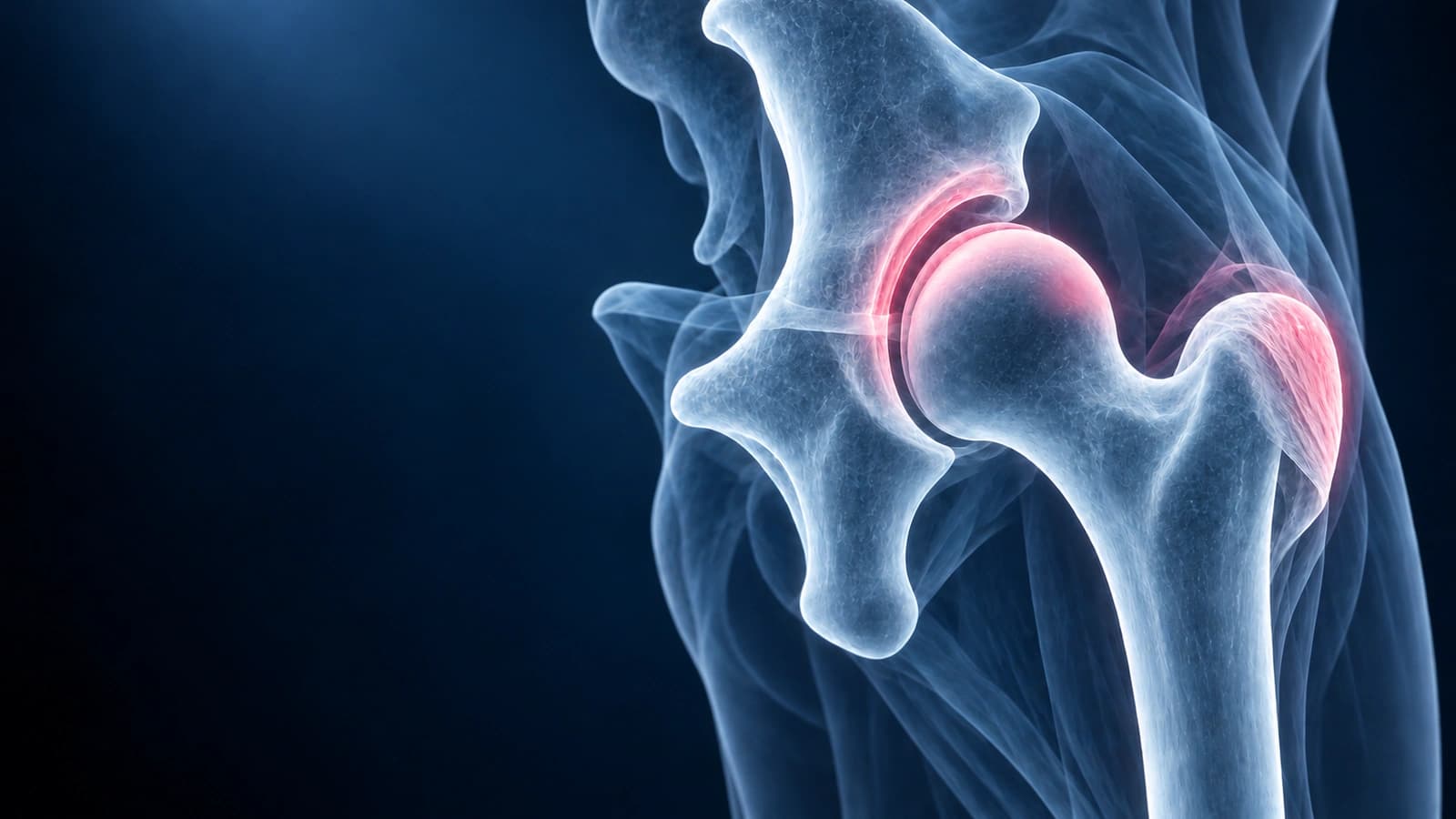
The hip is a ball-and-socket joint designed for stability and weight bearing. Its deep bony architecture provides inherent stability, but the surrounding labrum, cartilage, tendons, and bursae are susceptible to injury from both athletic activity and degenerative processes. Understanding common hip conditions helps patients navigate diagnosis and make informed treatment decisions.
Labral Tear
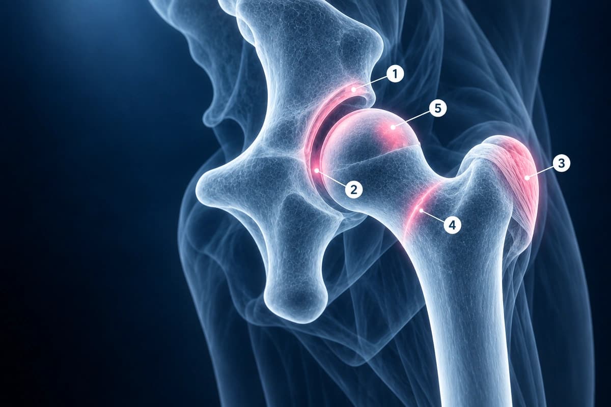
The acetabular labrum is a ring of fibrocartilage that deepens the hip socket and provides a suction seal for joint stability. Labral tears cause groin pain, clicking, catching, and stiffness, often worsened by prolonged sitting or hip flexion activities. They commonly result from femoroacetabular impingement (FAI), trauma, or hip dysplasia. MRI with arthrogram is the gold standard for diagnosis. For conservative management options, see our article on labral tear healing without surgery.
Femoroacetabular Impingement (FAI)
FAI occurs when abnormal bone morphology of the femoral head or acetabulum causes premature contact during hip motion. Cam-type impingement involves an aspherical femoral head, while pincer-type involves excessive acetabular coverage. Most patients have a combination of both (mixed type). FAI leads to labral tears and cartilage damage over time and is a major risk factor for early hip osteoarthritis. Activity modification and physiotherapy are first-line treatments, with hip arthroscopy for refractory cases. Learn how these morphology changes appear on MRI in our hip MRI reading guide.
Greater Trochanteric Bursitis
Now more accurately called greater trochanteric pain syndrome, this condition involves inflammation of the bursa and often coexistent gluteus medius or minimus tendinopathy. It causes lateral hip pain that is worse with lying on the affected side, climbing stairs, and prolonged walking. Treatment includes activity modification, physical therapy focusing on hip abductor strengthening, and corticosteroid injection for refractory pain. True gluteal tendon tears may require surgical repair in severe cases.
Stress Fracture
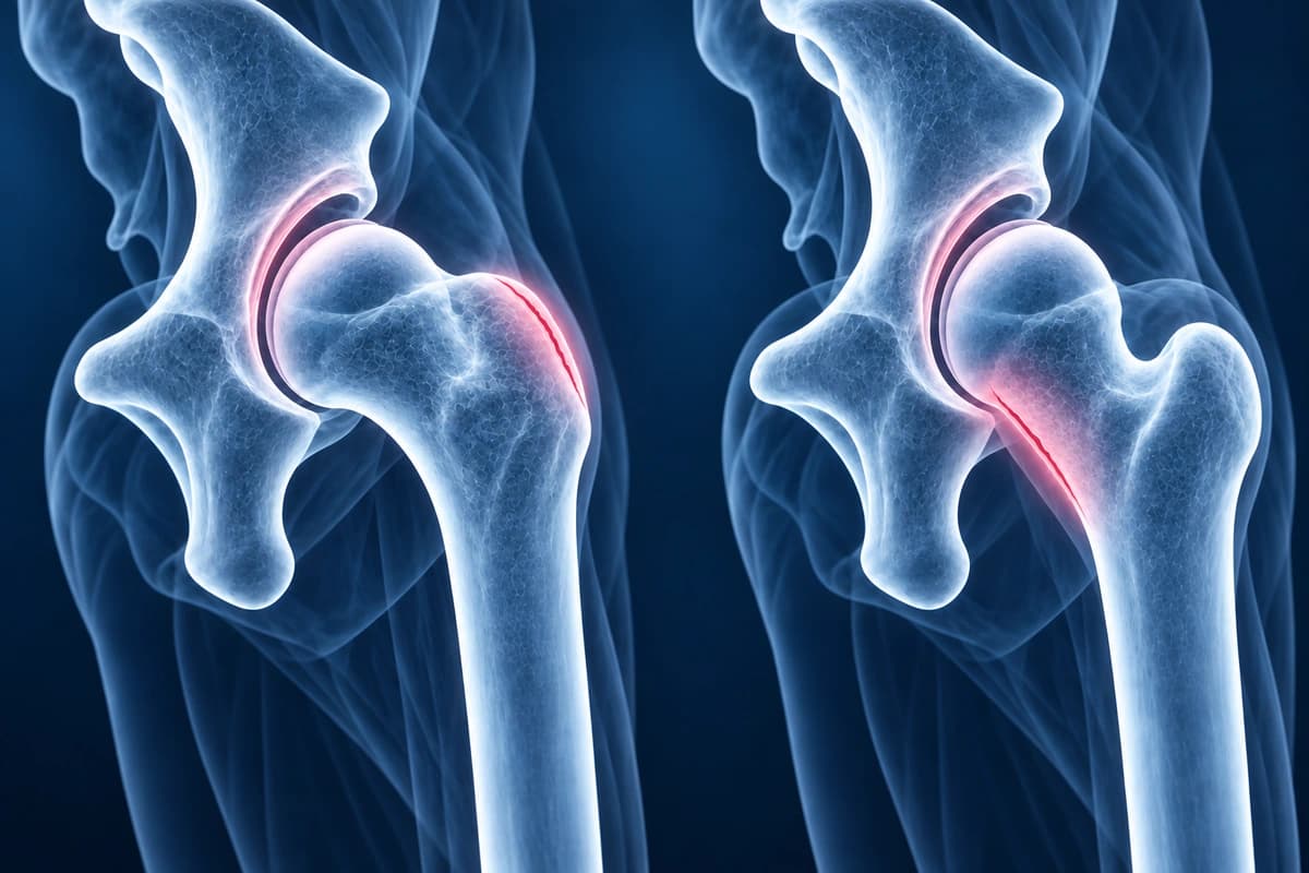
Hip stress fractures most commonly affect the femoral neck, particularly in runners, military recruits, and patients with metabolic bone disease. They present with groin pain that worsens with weight-bearing activity. Femoral neck stress fractures are classified as tension-side (lateral, higher surgical risk) or compression-side (medial, generally treated conservatively). MRI is essential for early detection as X-rays may be normal for weeks. Prompt diagnosis prevents progression to complete fracture.
Avascular Necrosis (AVN)
AVN of the femoral head occurs when blood supply to the bone is disrupted, causing bone death and eventual joint collapse. Risk factors include corticosteroid use, alcohol abuse, sickle cell disease, and hip trauma. Early stages are asymptomatic and detectable only on MRI, making screening important for high-risk patients. Early AVN may be managed with core decompression surgery, while advanced collapse typically requires total hip replacement.
Frequently Asked Questions
What causes hip pain in young adults?
The most common causes of hip pain in young adults are labral tears, FAI, hip flexor tendinitis, and stress fractures. Unlike older adults where osteoarthritis dominates, young patients more often have structural or overuse conditions that are highly treatable when diagnosed early.
Do I need an MRI for hip pain?
MRI is recommended when hip pain persists beyond 4 to 6 weeks despite conservative measures, when there is suspicion for labral tear or stress fracture, or when the diagnosis is unclear. X-rays should be obtained first to evaluate for arthritis, fractures, and bony morphology. MRI with arthrogram is preferred for suspected labral tears.
Sources
- Ganz R, Parvizi J, Beck M, et al. Femoroacetabular impingement: a cause for osteoarthritis of the hip. Clin Orthop Relat Res. 2003;(417):112-120.
- Long SS, Surrey DE, Nazarian LN. Sonography of greater trochanteric pain syndrome and the rarity of primary bursitis. AJR Am J Roentgenol. 2013;201(5):1083-1086.
- Fullerton LR Jr, Snowdy HA. Femoral neck stress fractures. Am J Sports Med. 1988;16(4):365-377.
- Moya-Angeler J, Gianakos AL, Villa JC, et al. Current concepts on osteonecrosis of the femoral head. World J Orthop. 2015;6(8):590-601.
Related Articles
Understand your hip MRI report including labrum evaluation, cam and pincer morphology, cartilage assessment, and AVN detection.
Find out if hip labral tears can resolve without surgery, the role of physiotherapy, and surgical options when conservative care fails.
Related Conditions
Understand your imaging before your next appointment
MRI, X-ray or CT from a CD, folder or ZIP. A PDF report or photos of the films work too.
Free to open and preview, no account needed. An optional AI report from $10 explains your images in plain language, with questions for your doctor.
Your raw files stay on this device. Only rendered images and minimal context are sent when you request an AI report.
Medical Disclaimer: This page is for informational and educational purposes only. It does not constitute medical advice, diagnosis, or treatment. AI-generated analysis may contain errors. Always consult a qualified healthcare professional for medical decisions. Full Disclaimer