
Hip AVN: Ficat & ARCO Staging Explained on MRI
Avascular necrosis of the femoral head — Ficat I-IV and ARCO 1-4 staging, MRI signs (double-line sign, crescent sign, collapse), and joint-preserving vs replacement options.
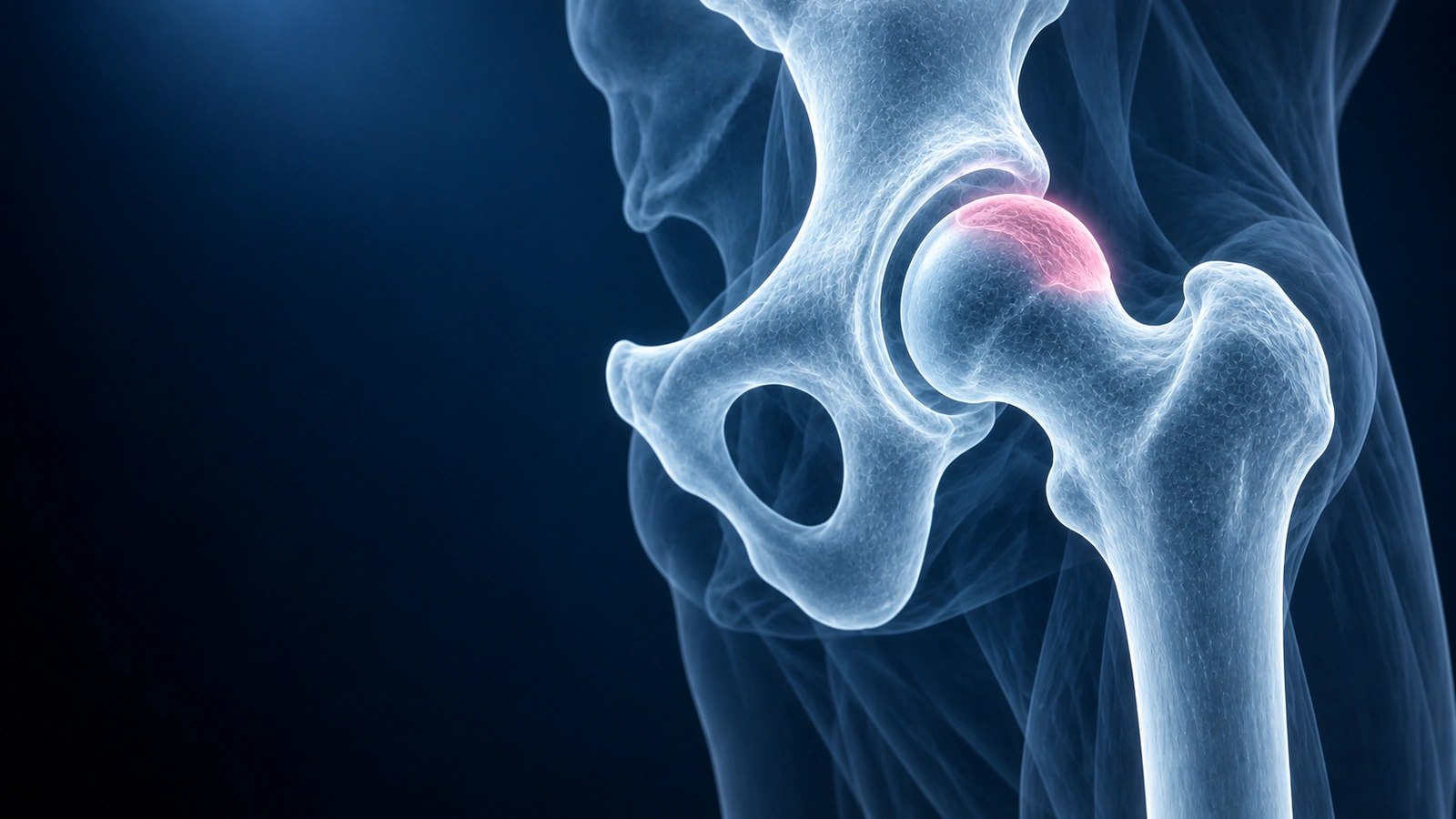
Avascular necrosis (AVN) of the femoral head is a condition in which blood supply to the bone is lost, causing bone cells to die. Without treatment, the weakened bone gradually collapses under the weight of the body, destroying the hip joint. AVN can affect anyone but is particularly common in patients who have taken corticosteroids, those with excessive alcohol use, and those who have sustained a hip fracture or dislocation. It strikes people in their 30s and 40s as often as older adults — making timely diagnosis and staging essential.
MRI detects AVN years before any changes appear on plain X-ray, making it the single most important imaging tool for this condition. Once AVN is confirmed, staging determines whether the goal is joint preservation or joint replacement. This article explains both the Ficat and ARCO staging systems, the key MRI findings at each stage, and which treatments are appropriate at each level of severity.
Why Staging Matters
AVN follows a predictable but highly variable course. In the earliest stages, the necrotic segment of bone is still structurally intact — it has not collapsed, and the joint surface is still smooth. Joint-preserving procedures such as core decompression, vascularized bone grafting, or protected weight-bearing can slow or halt progression and allow patients to keep their own hip for decades. Once the femoral head collapses and the cartilage is destroyed, joint preservation is no longer realistic, and total hip arthroplasty becomes the definitive treatment. Staging makes this distinction precise and reproducible so that surgeons and patients can plan together.
Ficat Stage I: Pre-Radiographic Disease
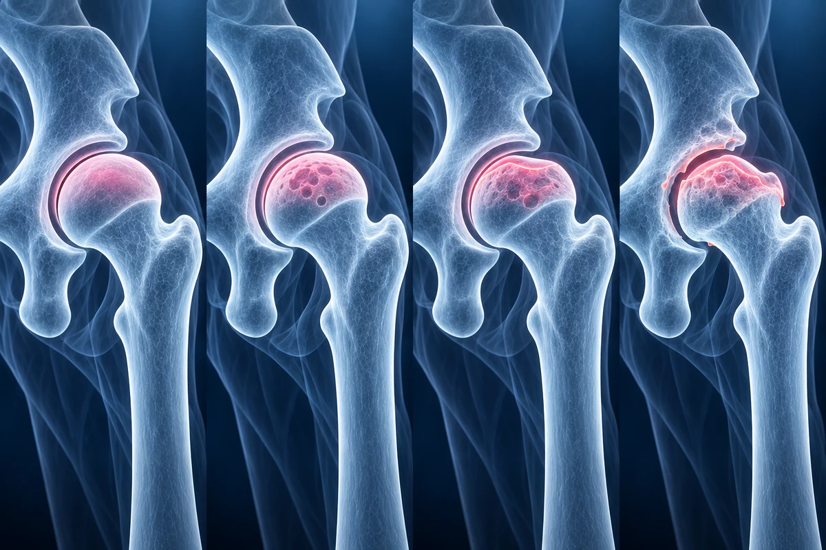
In Ficat Stage I, plain X-rays appear completely normal, yet the patient typically has hip or groin pain and the necrotic process is already underway. MRI is the only imaging modality that reliably detects this stage. The characteristic MRI findings are bone marrow edema — seen as increased signal on T2-weighted fat-suppressed sequences — and the double-line sign on T2-weighted images. The double-line sign consists of an inner hyperintense rim of granulation tissue surrounded by a hypointense band of reactive sclerosis; when present, it is pathognomonic for AVN, meaning no other condition produces this exact pattern. Identifying Stage I disease is critical because it represents the best window for joint-preserving treatment.
Ficat Stage II: Radiographic Changes Without Collapse
Ficat Stage II is defined by visible changes on plain X-ray — including areas of sclerosis, cyst formation, or mixed density within the femoral head — while the overall spherical contour of the femoral head remains intact and there is no collapse. On MRI, the necrotic segment is clearly demarcated and typically shows low T1 signal and a well-defined reactive interface. The size of the necrotic lesion matters enormously at this stage: small lesions in the medial femoral head carry a low risk of collapse, while large lesions occupying more than one-third of the femoral head surface are at high risk of progressing regardless of treatment.
Ficat Stage III: Subchondral Collapse and the Crescent Sign
Stage III marks a pivotal and often irreversible threshold: the subchondral bone beneath the dead segment fractures, producing the crescent sign — a thin radiolucent line paralleling the articular surface on X-ray or a subchondral fracture line on MRI. The articular cartilage itself may still be intact at this stage, but the structural support beneath it has failed. Once the crescent sign is visible, collapse of the femoral head typically follows within weeks to months without intervention. The amount of depression (measured in millimetres on coronal MRI) guides surgical decisions: small collapse under 2 mm may still be approached with joint-preserving surgery; collapse greater than 4 mm generally predicts a poor result with anything other than total hip arthroplasty.
Ficat Stage IV: Acetabular Involvement and End-Stage Arthritis
In Ficat Stage IV, the collapsed and flattened femoral head begins to damage the acetabular cartilage. X-rays show joint space narrowing, acetabular sclerosis, and often osteophyte formation — the full picture of secondary osteoarthritis. MRI confirms the extent of cartilage loss on both the femoral and acetabular sides. At this stage, the patient experiences severe pain, markedly limited range of motion, and significant functional disability. Total hip replacement is the standard treatment and reliably restores function and eliminates pain.
ARCO 1–4: The Modern Equivalent
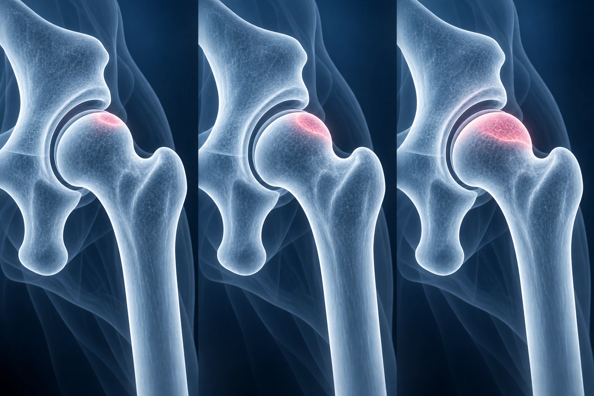
The Association Research Circulation Osseous (ARCO) classification was developed to standardise AVN staging internationally and to add lesion-size substaging that the Ficat system lacks. ARCO 1 corresponds to Ficat Stage I — MRI-detectable disease with normal X-rays — and is further subdivided by lesion size into 1A (less than 15% of femoral head), 1B (15–30%), and 1C (greater than 30%). ARCO 2 corresponds to Ficat Stage II, with visible X-ray changes but no collapse. ARCO 3 corresponds to Ficat Stage III, defined by subchondral fracture and crescent sign, with subclassification by the degree of depression in millimetres. ARCO 4 corresponds to Ficat Stage IV with acetabular involvement. Most major orthopaedic centres now use ARCO staging because the lesion-size information directly informs treatment strategy: the same stage may carry a very different prognosis depending on whether the lesion is small or large.
Treatment by Stage
For Stage I and early Stage II (small lesions, ARCO 1A–2A), the approach is joint preservation. Protected weight-bearing reduces mechanical loading on the at-risk segment. Bisphosphonates, by reducing bone turnover, may slow the expansion of the necrotic area, though evidence is stronger for symptom relief than for halting progression. Core decompression — drilling one or more channels into the necrotic segment through the femoral neck — decompresses the elevated intraosseous pressure and creates a channel for revascularisation. Adding concentrated bone marrow aspirate (stem cells) to the decompression tunnel has shown improved outcomes in several prospective trials, particularly in Stage I and Stage IIA disease.
For Stage IIb–III with larger lesions and early collapse, vascularised fibular grafting — where a segment of the patient's own fibula with its blood supply is inserted into the femoral head to provide structural support and new vascularity — offers the best chance of preserving the native joint. Free vascularised iliac crest grafts are an alternative. These procedures are technically demanding and performed at specialist centres. When collapse exceeds 4 mm or the patient is older with lower functional demands, total hip arthroplasty is often a more reliable choice even at Stage III.
For Stage IV, total hip arthroplasty is the definitive treatment. Modern implants provide excellent pain relief and restore function to near-normal in most patients. Younger patients with AVN who undergo hip replacement should discuss implant choice and activity restrictions with their surgeon, as the goal is a prosthesis that will last many decades. For more on how AVN appears on MRI and what the AI analysis looks for, see the AVN condition page. For general guidance on reading a hip MRI report, see our hip MRI reading guide.
Key Takeaways
- MRI detects AVN years before X-ray changes — Ficat I and ARCO 1 are invisible on plain radiographs
- The double-line sign on T2-weighted MRI is pathognomonic for AVN and is the key Stage I finding
- The crescent sign marks subchondral fracture (Stage III) and signals that collapse is imminent without intervention
- Lesion size within each stage strongly predicts collapse risk — ARCO substaging (A, B, C) encodes this information
- Stage I–IIa disease with small lesions is best treated with core decompression ± stem cells to preserve the native joint
- Stage IV disease with acetabular involvement requires total hip arthroplasty, which reliably restores function and eliminates pain
Frequently Asked Questions
Can early-stage AVN reverse on its own?
Spontaneous reversal is rare but does occur in a subset of Stage I cases, particularly those with very small lesions or those in which the underlying cause (such as corticosteroid use or alcohol) has been eliminated. Transient bone marrow oedema of the hip — sometimes called transient osteoporosis — can mimic early AVN on MRI and does resolve spontaneously; distinguishing the two requires careful MRI interpretation. True AVN with the double-line sign almost never reverses without intervention, and watchful waiting without treatment is only appropriate for genuinely small lesions where the risk of progression is accepted as low.
What exactly is the double-line sign on MRI?
The double-line sign is seen on T2-weighted MRI sequences and consists of two concentric bands at the reactive interface between healthy and necrotic bone. The inner band appears bright (hyperintense) because it represents vascular granulation tissue — the body's attempt to revascularise the dead bone. The outer band appears dark (hypointense) because it represents reactive sclerosis and fibrous tissue. This two-layer appearance is unique to avascular necrosis and, when present, allows the diagnosis to be made with high confidence without a biopsy.
Does core decompression actually work?
Core decompression is most effective when performed at Stage I and small Stage IIA disease. Multiple studies show that approximately 70–80% of Stage I hips and 60–70% of Stage IIA hips treated with core decompression do not progress to collapse over 5 years. Results deteriorate significantly with larger lesions or more advanced staging. Adding concentrated bone marrow aspirate (containing stem cells) to the decompression channel appears to improve outcomes further in several randomised trials. It is not effective for Stage III disease with established collapse.
Do I need a hip replacement at Stage III?
Not necessarily. The decision at Stage III depends on several factors: the degree of collapse (less than 2 mm is more amenable to joint-preserving surgery than greater than 4 mm), the size of the lesion, patient age, activity demands, and the availability of specialist vascularised grafting. In younger patients with early Stage III and small lesions, vascularised fibular grafting can delay or prevent the need for replacement by 10 or more years. In older patients or those with larger collapse, total hip replacement delivers faster and more reliable relief and is often the pragmatic choice. Your surgeon will measure the collapse on coronal MRI as part of surgical planning.
What causes avascular necrosis of the hip?
The two most common causes are corticosteroid use and excessive alcohol consumption, together accounting for roughly 90% of non-traumatic cases. Corticosteroids cause fat emboli and impair bone cell survival; even short high-dose courses can trigger AVN in susceptible individuals. Alcohol promotes fat accumulation in bone and disrupts the lipid metabolism of bone cells. Traumatic AVN follows a displaced femoral neck fracture or hip dislocation that physically disrupts the retinacular blood vessels supplying the femoral head. Sickle cell disease causes AVN through sickling of red cells in the small vessels of the femoral head. Other causes include systemic lupus, Gaucher disease, radiation therapy, decompression sickness in divers, and hyperlipidaemia. In a minority of cases no cause is found and the AVN is classified as idiopathic.
Related Articles
Understand your hip MRI report including labrum evaluation, cam and pincer morphology, cartilage assessment, and AVN detection.
Learn about common hip conditions including labral tears, femoroacetabular impingement, bursitis, stress fractures, and avascular necrosis.
Related Conditions
Ready to analyze your imaging? Upload your MRI or X-ray for AI-powered analysis
Upload your MRI or X-ray DICOM files for private, AI-powered analysis. 4 models analyze independently — raw scan files stay in your browser.
Start AnalysisMedical Disclaimer: This page is for informational and educational purposes only. It does not constitute medical advice, diagnosis, or treatment. AI-generated analysis may contain errors. Always consult a qualified healthcare professional for medical decisions. Full Disclaimer