
FAI Cam vs Pincer Impingement: MRI Differences Explained
Cam, pincer, and mixed femoroacetabular impingement on MRI — alpha angle, lateral center-edge angle, labral tear patterns, and treatment differences.
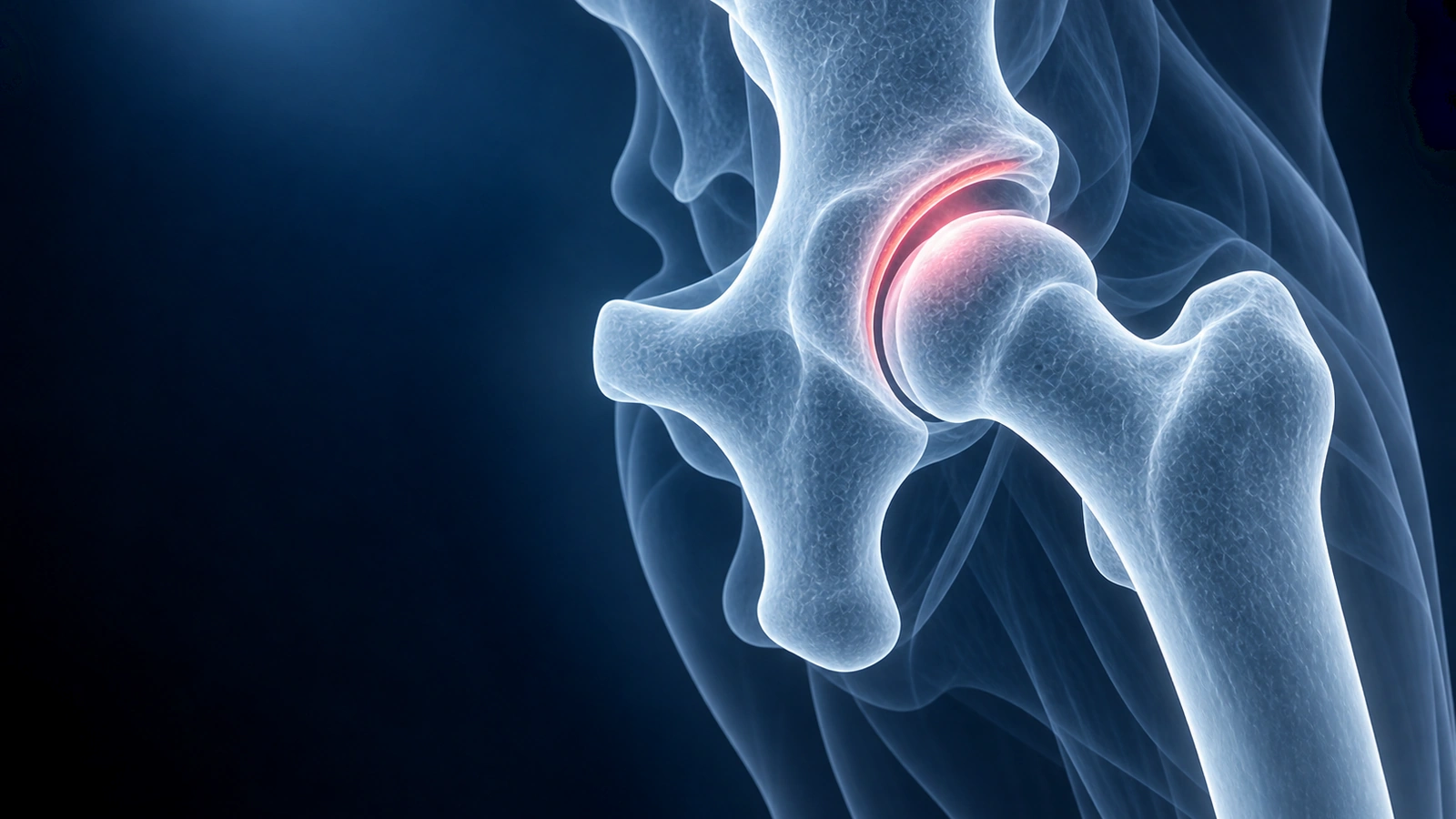
Femoroacetabular impingement (FAI) occurs when abnormal contact between the femoral head-neck junction and the acetabular rim generates repeated mechanical stress during hip motion. This abnormal contact damages the labrum and articular cartilage over time, leading to pain, functional limitation, and — if left untreated — early hip osteoarthritis.
FAI is classified into three morphological subtypes: cam, pincer, and mixed. Each has a distinct structural cause, characteristic MRI appearance, and different pattern of labral and cartilage injury. MRI and radiographs are complementary — plain X-rays quantify bony morphology under load, while MRI reveals soft-tissue damage, labral status, cartilage integrity, and the precise anatomy of the impingement zone. For a broader introduction to hip impingement, see our dedicated condition page.
Cam Impingement (Femoral-sided)
Cam impingement arises from an aspherical femoral head-neck junction — the normal concave waist is replaced by a convex or flat bony prominence. During hip flexion and internal rotation, this excess bone engages the acetabular rim and shears the anterosuperior labrum and adjacent cartilage from inside out.
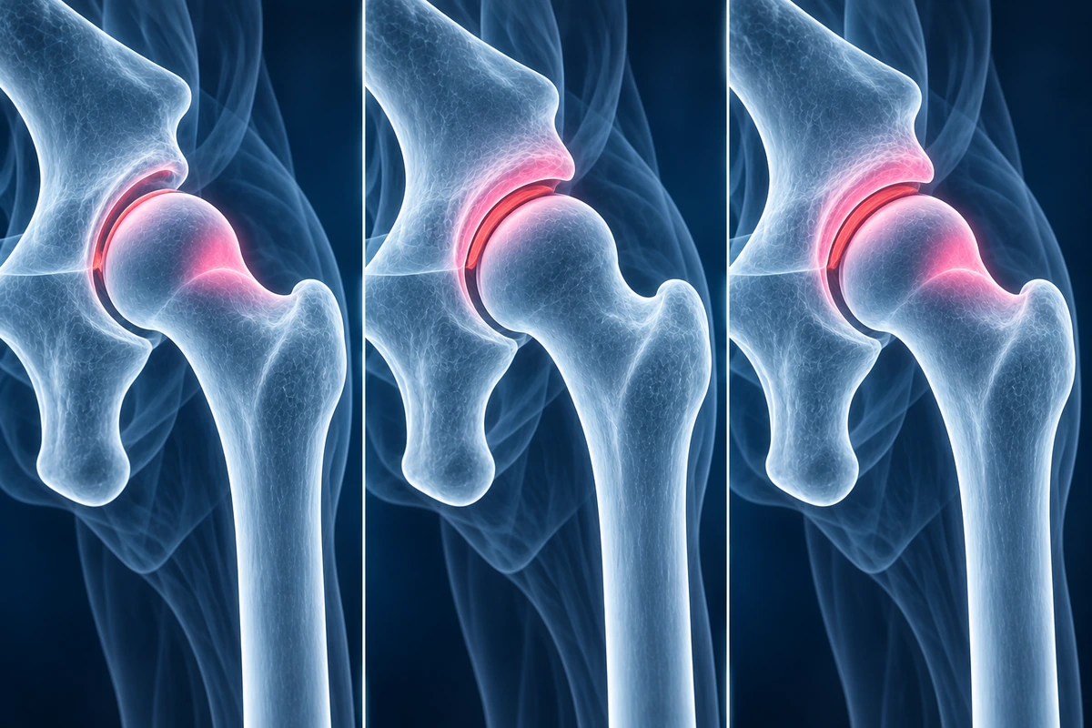
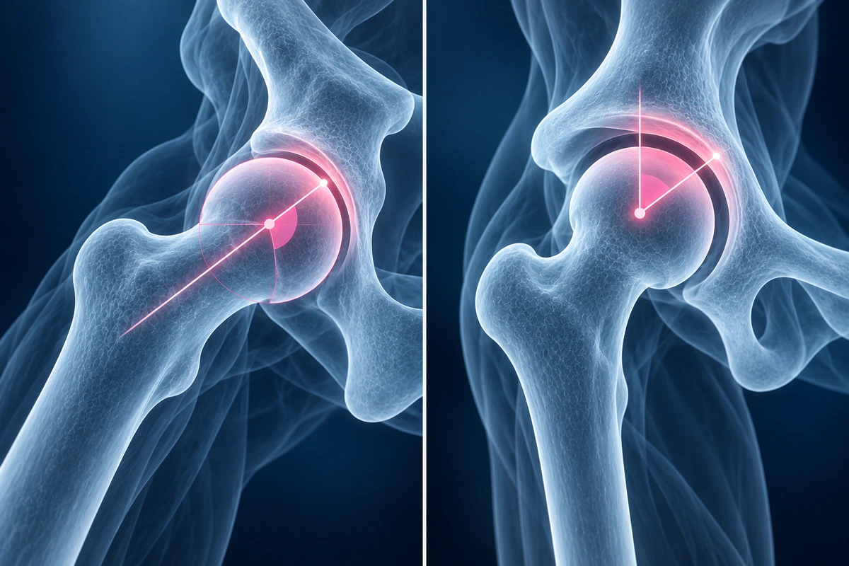
Cam deformities are most prevalent in young athletic males who performed high-impact sport during skeletal growth — the deformity is believed to develop at the proximal femoral physis under repetitive load. On MRI, cam morphology appears as a loss of the normal femoral head-neck offset on oblique axial sequences, often with a discrete bony bump at the anterosuperior head-neck junction. The alpha angle, measured on a radial or oblique axial image through the axis of the femoral neck, quantifies the asphericity. An alpha angle greater than 55 degrees is the widely accepted threshold for abnormal cam morphology, although some studies use 60 degrees. 3T MRI and radial reformats that sample every clock position around the head-neck junction improve alpha angle accuracy and detect subtle off-axis cam lesions that a standard axial sequence can miss.
Pincer Impingement (Acetabular-sided)
Pincer impingement results from acetabular over-coverage of the femoral head. The acetabular rim contacts the femoral neck directly and repetitively, crushing the labrum between the two bony surfaces. Three structural variants produce this over-coverage: general coxa profunda or protrusio acetabuli (the socket is globally too deep), focal anterior over-coverage from acetabular retroversion, and excessive lateral coverage reflected by a high lateral center-edge angle.
Pincer morphology is more common in middle-aged women than in men and often presents with more gradual, activity-related groin pain rather than the acute sporting episode typical of cam injuries. On MRI and radiographs, the lateral center-edge angle (LCEA) measured on a standing AP pelvis or coronal MRI greater than 40 degrees indicates lateral over-coverage. Acetabular retroversion produces the crossover sign on radiographs — the anterior acetabular wall projects lateral to the posterior wall. On MRI, the anterior wall can be measured to confirm focal anterior over-coverage. MR arthrography outlines the labrum with injected contrast and is the most sensitive technique for diagnosing the labral tear patterns that pincer impingement generates.
Mixed FAI (Most Common Presentation)
Most symptomatic FAI cases — approximately 85 percent in surgical series — show both cam and pincer morphology simultaneously. The two deformities interact: cam asphericity generates shear forces at the anterosuperior rim while pincer over-coverage produces direct rim loading. The resulting labral and cartilage injury pattern reflects contributions from both mechanisms and tends to be more severe than either type in isolation.
On MRI, mixed FAI shows an elevated alpha angle together with increased LCEA or evidence of retroversion. The labrum is typically torn at the anterosuperior position, and cartilage damage extends over a wider arc than in pure cam or pure pincer disease. Surgical planning for mixed FAI must address both the femoral-sided cam resection and the acetabular-sided rim trimming or periacetabular correction to prevent recurrent impingement.
Key MRI Measurements
- Alpha angle greater than 55 degrees on radial or oblique axial MRI: abnormal cam morphology; measured from the femoral neck axis to the point where the femoral head loses its sphericity
- Lateral center-edge angle greater than 40 degrees on coronal MRI or AP radiograph: excessive lateral acetabular coverage consistent with pincer over-coverage
- Anterior wall index and crossover sign: quantify focal anterior over-coverage from acetabular retroversion; best seen on false-profile radiograph and axial MRI
- Femoral head-neck offset ratio: a ratio below 0.17 on oblique axial MRI correlates with symptomatic cam impingement in clinical series
- 3T MRI with radial reformats: preferred protocol for alpha angle measurement because it samples the head-neck junction at every clock position, detecting off-axis cam deformities invisible on a single oblique axial slice
Labral and Cartilage Patterns
Cam impingement generates an inside-out shear force at the anterosuperior acetabular rim during flexion and internal rotation. The resulting labral tear is typically a detachment or delamination at the chondrolabral junction in the anterosuperior quadrant (12 to 2 o'clock position on axial arthrogram images). The adjacent acetabular cartilage peels away from bone in a wave-like sheet — the carpet sign on MR arthrography — which is highly specific for cam-pattern chondral damage.
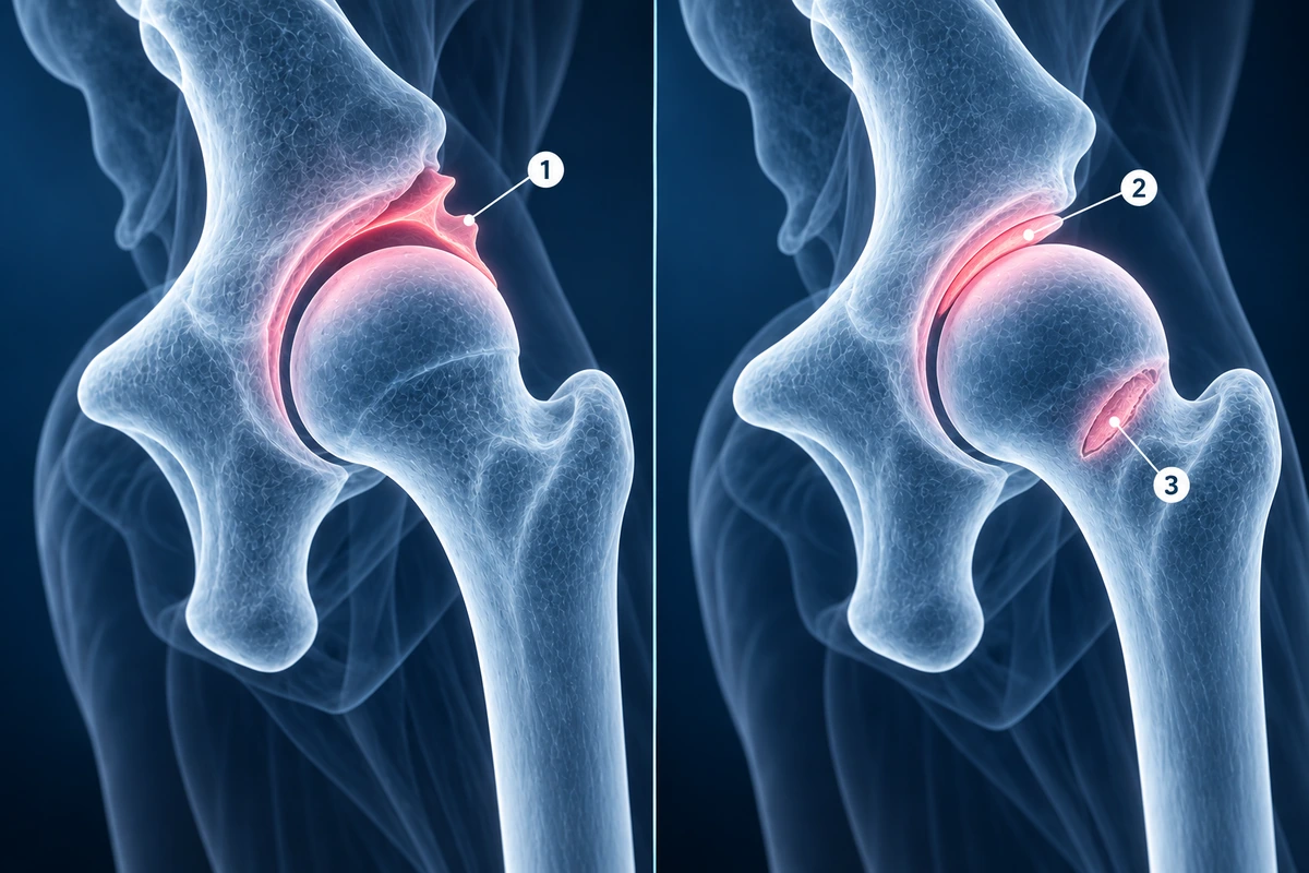
Pincer impingement compresses the labrum directly against the rim. The labrum degenerates and may ossify over time, and the initial cartilage damage is a narrow stripe at the anterosuperior acetabular rim directly beneath the impingement zone. A characteristic secondary finding is contre-coup chondral injury on the posteroinferior femoral head: as the pincer rim levers against the femoral neck, the posterior femoral head impacts the posteroinferior acetabulum, producing a small cartilage lesion remote from the primary impingement site. Identifying contre-coup injury on MRI strongly suggests pincer-pattern disease even when the labral tear itself is subtle. For a full guide to interpreting hip MRI sequences and measurements, see our article on how to read a hip MRI.
Treatment Implications
Identifying the FAI subtype on MRI directly shapes both non-operative and surgical management. Conservative treatment — physiotherapy targeting hip external rotator and core strength, activity modification to avoid terminal hip flexion, and anti-inflammatory medication — is the recommended first-line approach for all subtypes and succeeds in a meaningful proportion of patients, particularly those with mild morphological abnormality and no significant cartilage damage.
When surgery is indicated, cam FAI is treated by femoral osteochondroplasty — arthroscopic or open resection of the bony bump to restore femoral head sphericity and a normal alpha angle. Pincer FAI with acetabular retroversion may require arthroscopic rim trimming, labral refixation, and in more severe cases a periacetabular osteotomy (PAO) to reorient the entire socket. Mixed FAI combines both procedures. Labral tears are addressed simultaneously — repaired when the tissue quality permits, reconstructed with a graft when the labrum is deficient. Chondral damage identified on MRI, including the carpet sign of cam delamination and contre-coup pincer lesions, is treated with microfracture, chondroplasty, or cartilage restoration depending on lesion size and depth.
Key Takeaways
- Cam FAI: aspherical femoral head-neck junction (alpha angle greater than 55 degrees), most common in young athletic males, causes inside-out anterosuperior chondrolabral shear
- Pincer FAI: acetabular over-coverage (LCEA greater than 40 degrees or retroversion crossover sign), more common in middle-aged women, compresses the labrum against the rim
- Mixed FAI accounts for approximately 85 percent of symptomatic cases and shows both elevated alpha angle and increased acetabular coverage
- Carpet sign on MR arthrography — delamination of acetabular cartilage in a sheet — is highly specific for cam-pattern chondral injury
- Contre-coup chondral injury on the posteroinferior femoral head is characteristic of pincer impingement and can be identified on MRI
- 3T MRI with radial reformats and MR arthrography provides the most complete assessment of FAI morphology, labral tears, and cartilage damage for surgical planning
Frequently Asked Questions
Who typically develops cam versus pincer FAI?
Cam deformities are significantly more prevalent in males and in individuals who participated in high-impact sport — particularly football, ice hockey, and basketball — during adolescence. The repetitive loading of the proximal femoral physis during growth is thought to drive cam development. Pincer morphology is more evenly distributed between sexes but is more commonly symptomatic in middle-aged women, partly because acetabular retroversion has a higher prevalence in females. Mixed morphology is the most common presentation regardless of sex.
Is the 55-degree alpha angle threshold for cam FAI universally agreed upon?
No. The threshold is debated in the literature. Most studies use 55 degrees as the cutoff for abnormal cam morphology, but some groups use 60 degrees to improve specificity. Alpha angle also varies by the clock position at which it is measured — the highest value typically occurs at the 1 to 2 o'clock position on radial reformats rather than on a single oblique axial image. Because of this variability, the alpha angle should be interpreted alongside clinical symptoms, impingement tests, and other MRI findings rather than used as a binary diagnostic criterion in isolation.
Can FAI be present without causing symptoms?
Yes, and this is common. Population imaging studies show that cam and pincer morphology are prevalent in asymptomatic individuals — cam deformities are detectable in up to 25 percent of asymptomatic men in some series. The presence of FAI morphology on MRI or radiographs does not by itself establish a diagnosis of symptomatic FAI or indicate that surgery is warranted. Clinical correlation — groin pain reproduced by the FADIR test (flexion, adduction, internal rotation), activity-related symptoms, and a labral tear on MRI — is required before attributing a patient's symptoms to the morphological finding.
Does FAI always require surgery, or can physiotherapy resolve it?
Surgery is not inevitable. A structured physiotherapy programme addressing hip external rotator strength, core stability, and movement pattern retraining resolves symptoms satisfactorily in a proportion of patients, particularly those with mild morphological abnormality, intact cartilage, and no large labral tear. The UK FASHION trial (2018) found that hip arthroscopy was not significantly superior to personalised physiotherapy at 12 months for patient-reported outcomes, although surgical patients reported better function at some later timepoints. Current guidelines recommend completing at least three to six months of supervised physiotherapy before surgical referral in patients without severe cartilage damage.
Does untreated FAI lead to hip osteoarthritis?
Evidence supports a causal link between FAI morphology and early hip osteoarthritis, but the relationship is not deterministic. Longitudinal cohort studies show that hips with cam morphology and labral tears have higher rates of cartilage progression than morphologically normal hips over five to ten years. However, many individuals with FAI morphology never develop clinical osteoarthritis. Risk factors for progression include large alpha angle, significant chondral damage at presentation (particularly the carpet sign of full-thickness delamination), age over 40, and elevated body mass index. Successful surgical correction that restores normal morphology appears to slow cartilage progression, but long-term data beyond ten years are still limited.
Related Articles
Understand your hip MRI report including labrum evaluation, cam and pincer morphology, cartilage assessment, and AVN detection.
Find out if hip labral tears can resolve without surgery, the role of physiotherapy, and surgical options when conservative care fails.
Related Conditions
Ready to analyze your imaging? Upload your MRI or X-ray for AI-powered analysis
Upload your MRI or X-ray DICOM files for private, AI-powered analysis. 4 models analyze independently — raw scan files stay in your browser.
Start AnalysisMedical Disclaimer: This page is for informational and educational purposes only. It does not constitute medical advice, diagnosis, or treatment. AI-generated analysis may contain errors. Always consult a qualified healthcare professional for medical decisions. Full Disclaimer